The brain is the main regulator of the functions of any living organism, one of the elements. Until now, medical scientists are studying the features of the brain and discovering all of its incredible new possibilities. It is a very complex organ that connects our body with the external environment. The parts of the brain and their functions regulate all life processes. External receptors catch signals and inform any part of the brain about incoming stimuli (light, sound, tactile and many others). The response is immediate. Let's take a closer look at how our head processor works.
General description of the brain
The parts of the brain and their functions completely control our life processes. The human brain consists of 25 billion neurons. This incredible number of cells forms gray matter. The brain covers several membranes:
- soft;
- solid;
- spider web (cerebrospinal fluid circulates here).
CSF is a cerebrospinal fluid, in the brain it plays the role of a shock absorber, a protector from any shock force.
In both men and women, the brain is developed in exactly the same way, although its weight is different. More recently, controversy has subsided that brain weight plays a role in mental development and intellectual ability. The conclusion is unambiguous - it is not so. The weight of the brain is approximately 2% of the total weight of a person. In men, its weight is on average 1 370 g, and in women - 1 240 g. The functions of the human brain departments are developed in a standard way, vital activity depends on them. Mental abilities depend on quantitative connections created in the brain. Every brain cell is a neuron that generates and transmits impulses.
The cavities inside the brain are called the ventricles. The cranial paired nerves go to different departments.
Functions of the parts of the brain (table)
Each part of the brain does its job. The table below demonstrates this clearly. The brain, like a computer, clearly performs its tasks, receiving commands from the outside world.
The functions of the parts of the brain, the table reveals schematically and succinctly.
Below we will consider the parts of the brain in more detail.
Structure
The picture shows how the brain works. Despite this, the most significant part is occupied, all parts of the brain and their functions play a huge role in the work of the body. There are five main divisions:
- final (80% of the total mass);
- posterior (bridge and cerebellum);
- intermediate;
- oblong;
- average.
At the same time, the brain is divided into three main parts: the brain stem, the cerebellum, and the two cerebral hemispheres.

Ultimate brain
It is impossible to briefly describe the structure of the brain. To understand the parts of the brain and their functions, it is necessary to closely study their structure.
The telencephalon extends from the frontal to the occipital bone. There are two cerebral hemispheres considered here: the left and the right. This section differs from others in the largest number of grooves and convolutions. The development and structure of the brain are closely linked. Experts have identified three types of bark:
- ancient (with the olfactory tubercle, the anterior perforated substance, the lunar podmozolic and the lateral podmozolic gyrus);
- old (with dentate gyrus - fascia and hippocambus);
- new (represents the rest of the crust).
The hemispheres are separated by a longitudinal groove, in its depth there is a vault and a corpus callosum, which connect the hemispheres. The corpus callosum itself is lined and belongs to the neocortex. The structure of the hemispheres is quite complex and resembles a multilevel system. The frontal, temporal, parietal and occipital lobes, subcortex and cortex are distinguished here. A huge number of functions are performed by the large hemispheres. It is worth noting that the left hemisphere commands the right side of the body, and the right, on the contrary, commands the left.
Bark
The superficial layer of the brain is the cortex, it is 3 mm thick and covers the hemispheres. The structure consists of vertical nerve cells with processes. The cortex also contains efferent and afferent nerve fibers, as well as neuroglia. The parts of the brain and their functions are discussed in the table, and what is the cortex? Its most complex structure has horizontal layering. The structure has six layers:
- outer pyramidal;
- outer granular;
- internal granular;
- molecular;
- inner pyramidal;
- with fusiform cells.
Each has a different width, density, shape of neurons. Vertical bundles of nerve fibers give the cortex a vertical striation. The area of the cortex is approximately 2,200 square centimeters, and the number of neurons here reaches ten billion.
Parts of the brain and their functions: cortex
The bark directs several specific bodily functions. Each share is responsible for its own parameters. Let's consider the functions associated with calving in more detail:
- temporal - controls the sense of smell and hearing;
- parietal - is responsible for taste and touch;
- occipital - vision;
- frontal - complex thinking, movement and speech.
Each neuron is in contact with other neurons, there are up to ten thousand contacts (gray matter). Nerve fibers are white matter. Some part unites the hemispheres of the brain. White matter includes three types of fibers:
- associative link in one hemisphere different cortical areas;
- commissural connect the hemispheres to each other;
- projection ones communicate with the lower formations, have analyzer paths.
Considering the structure and functions of the parts of the brain, it is necessary to emphasize the role of gray and white matter. The hemispheres have (gray matter) inside, their main function is to transmit information. A white matter is located between the cerebral cortex and the basal nuclei. There are four parts to this:
- between the grooves in the convolutions;
- in the outer places of the hemispheres;
- included in the inner capsule;
- located in the corpus callosum.
The white matter found here is formed by nerve fibers and connects the cortex of the gyrus with the underlying departments. form the subcortex of the brain.
Ultimate brain - directs all vital functions of the body, as well as the intellectual abilities of a person.

Diencephalon
The brain regions and their functions (see the table above) include the diencephalon. If you look in more detail, it should be said that it consists of the ventral and dorsal parts. The hypothalamus belongs to the ventral, the thalamus, metathalamus, and also the epithalamus to the dorsal.
The thalamus is a mediator that directs the received irritation to the hemispheres. It is often referred to as the "visual hillock". It helps the body to quickly adapt to changes in the external environment. The thalamus is connected to the cerebellum using the limbic system.
The hypothalamus controls vegetative functions. The influence goes through the nervous system, and, of course, the endocrine glands. Regulates the work of the endocrine glands, controls the metabolism. The pituitary gland is located just below it. Body temperature, cardiovascular and digestive systems are regulated. The hypothalamus also governs our eating and drinking behavior, regulates wakefulness and sleep.
Rear
The hindbrain includes the pons located in front and the cerebellum, which is located behind. Studying the structure and functions of the parts of the brain, let us take a closer look at the structure of the pons: the dorsal surface is overlapped by the cerebellum, the ventral is represented by a fibrous structure. The fibers are directed laterally in this section. On each side of the bridge, they extend to the cerebellar middle leg. In appearance, the bridge resembles a thickened white ridge located above the medulla oblongata. The nerve roots extend into the bulbar-bridge groove.
The structure of the posterior bridge: in the frontal section, it can be seen that the section of the anterior (large ventral) and posterior (small dorsal) parts consists. Between them, the border is a trapezoidal body, the transverse thick fibers of which are ranked as the auditory pathway. Conductive function is completely dependent on the hindbrain.
Cerebellum (small brain)
The table "Division of the brain, structure, function" indicates that the cerebellum is responsible for the coordination and movement of the body. This section is located at the back of the bridge. The cerebellum is often referred to as the "small brain". It occupies the posterior cranial fossa, covers the rhomboid. The mass of the cerebellum is from 130 to 160 g. Above are the large hemispheres, which are separated by a transverse slit. The lower part of the cerebellum is adjacent to the medulla oblongata.
Two hemispheres are distinguished here, the lower, upper surface and the worm. The border between them is called a horizontal deep slot. Many cracks cut the surface of the cerebellum, between them are thin convolutions (rollers). Between the grooves there are groups of convolutions, divided into lobules, they represent the lobes of the cerebellum (posterior, clumpy-nodular, anterior).
The cerebellum contains both gray and gray located in the periphery, forms a cortex with molecular and pear-shaped neurons, and a granular layer. Under the bark there is a white substance that penetrates into the convolutions. In the white matter there are inclusions of gray (its nuclei). In cross-section, this ratio is similar to a tree. Those who know the structure of the human brain, the functions of its parts, will easily answer that the cerebellum is the regulator of the coordination of movements of our body.

Midbrain
The midbrain is located in the area of the anterior pons and goes to the papillary bodies, as well as to the optic tracts. Here, clusters of nuclei are highlighted, which are called the hillocks of the quadruple. The structure and functions of the parts of the brain (table) indicate that this department is responsible for latent vision, an orientation reflex, gives orientation to reflexes to visual and sound stimuli, and also maintains muscle tone in the human body.
Medulla oblongata: stem part
The medulla oblongata is a natural extension of the spinal cord. That is why the structure has a lot in common. This becomes especially clear if we examine in detail the white matter. It is represented by short and long nerve fibers. The gray matter is represented here in the form of nuclei. The parts of the brain and their functions (the table is presented above) indicates that the medulla oblongata controls our balance, coordination, regulates metabolism, controls respiration and blood circulation. It is also responsible for such important reflexes of our body as sneezing and coughing, vomiting.
The brainstem is subdivided into the hindbrain and midbrain. The trunk is called the middle, oblong, bridge and diencephalon. Its structure is descending and ascending paths connecting the trunk with the spinal cord and brain. In this part, heartbeat, breathing, articulate speech are monitored.
The brain is the main controlling organ of the central nervous system (CNS); a large number of specialists in various fields, such as psychiatry, medicine, psychology and neurophysiology, have been working on the study of its structure and functions for over 100 years. Despite a good study of its structure and components, there are still many questions about the work and processes taking place every second.
The brain belongs to the central nervous system and is located in the cranial cavity. Outside, it is reliably protected by the bones of the skull, and inside it is enclosed in 3 shells: soft, arachnoid and hard. Cerebrospinal fluid circulates between these membranes - cerebrospinal fluid, which serves as a shock absorber and prevents shaking of this organ in minor injuries.
The human brain is a system consisting of interconnected departments, each part of which is responsible for performing specific tasks.
It is not enough to briefly describe the brain to understand the functioning, therefore, to understand how it works, you first need to study its structure in detail.
What is the brain responsible for?


This organ, like the spinal cord, belongs to the central nervous system and plays the role of an intermediary between the environment and the human body. With its help, self-control, reproduction and memorization of information, figurative and associative thinking, and other cognitive psychological processes are carried out.
According to the teachings of Academician Pavlov, the formation of thought is a function of the brain, namely the cerebral cortex, which are the highest organs of nervous activity. The cerebellum, the limbic system and some parts of the cerebral cortex are responsible for different types of memory, but since memory is different, it is impossible to isolate any specific area responsible for this function.
He is responsible for the management of the autonomic vital functions of the body: respiration, digestion, endocrine and excretory systems, control of body temperature.
To answer the question of what function the brain performs, first it should be conditionally divided into sections.
Experts distinguish 3 main parts of the brain: anterior, middle and rhomboid (posterior) section.
- The front one performs higher psychiatric functions, such as the ability to cognize, the emotional component of a person's character, his temperament and complex reflex processes.
- The middle one is responsible for sensory functions and the processing of information received from the organs of hearing, sight and touch. The centers located in it are able to regulate the degree of pain sensations, since the gray matter, under certain conditions, is capable of producing endogenous opiates that raise or lower the pain threshold. It also plays the role of a conductor between the cortex and the underlying departments. This part controls the body through various innate reflexes.
- The rhomboid or posterior section is responsible for muscle tone, body coordination in space. Through it, a targeted movement of various muscle groups is carried out.
The structure of the brain cannot be simply briefly described, since each of its parts includes several departments, each of which performs specific functions.
What does the human brain look like?


Anatomy of the brain is a relatively young science, since for a long time it was banned due to laws prohibiting the opening and examination of organs and the head of a person.
The study of the topographic anatomy of the brain region in the head area is necessary for accurate diagnosis and successful treatment of various topographic anatomical disorders, for example: skull injuries, vascular and oncological diseases. To imagine what a human GM looks like, you first need to study their appearance.
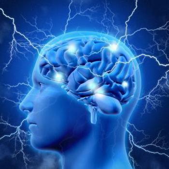
In appearance, GM is a yellowish gelatinous mass, enclosed in a protective shell, like all organs of the human body, they consist of 80% water.
The large hemispheres occupy almost the volume of this organ. They are covered with gray matter or bark - the highest organ of human neuropsychic activity, and inside - of white matter, consisting of the processes of nerve endings. The surface of the hemispheres has a complex pattern, due to the convolutions and ridges going in different directions between them. According to these convolutions, it is customary to divide them into several departments. It is known that each of the parts performs specific tasks.
In order to understand what a person's brains look like, it is not enough to examine their appearance. There are several study methods that can help you view the inside of the brain in a section.
- Sagittal incision. It is a longitudinal section that goes through the center of the human head and divides it into 2 parts. It is the most informative research method, with its help various diseases of this organ are diagnosed.
- The frontal section of the brain looks like a cross section of the large lobes and allows you to see the fornix, hippocampus and corpus callosum, as well as the hypothalamus and thalamus, which control vital body functions.
- Horizontal section. Allows you to consider the structure of this organ in the horizontal plane.
Brain anatomy, like the anatomy of the human head and neck, is a rather difficult subject to study for a number of reasons, including the fact that a large amount of material and good clinical training are required to describe them.
How the human brain works
Scientists around the world are studying the brain, its structure and the functions that it performs. Over the past few years, many important discoveries have been made, however, this part of the body remains not fully understood. This phenomenon is explained by the difficulty of studying the structure and functions of the brain separately from the cranium.
In turn, the structure of the brain structures determines the functions that are performed by its departments.
It is known that this organ consists of nerve cells (neurons) interconnected by bundles of filamentous processes, but how their interaction occurs simultaneously as a single system is still not clear.
A diagram of the structure of the brain, based on the study of a sagittal section of the cranium, will help to investigate the departments and membranes. In this figure, you can see the cortex, the medial surface of the cerebral hemispheres, the structure of the trunk, cerebellum and corpus callosum, which consists of a ridge, trunk, knee and beak.
GM is reliably protected outside by the bones of the skull, and inside by 3 meninges: hard arachnoid and soft. Each of them has its own device and performs specific tasks.
- The deep soft membrane covers both the spinal cord and the brain, while entering all the cracks and grooves of the cerebral hemispheres, and in its thickness are the blood vessels that feed this organ.
- The arachnoid membrane is separated from the first by a subarachnoid space filled with cerebrospinal fluid (cerebrospinal fluid); it also contains blood vessels. This shell consists of connective tissue, from which the filamentous branching processes (strands) depart, they are woven into the soft shell and their number increases with age, thereby strengthening the connection. Between them. The villous outgrowths of the arachnoid membrane protrude into the lumen of the sinuses of the dura mater.
- The hard shell, or pachymeninx, consists of a connective tissue substance and has 2 surfaces: the upper one, saturated with blood vessels, and the inner one, which is smooth and shiny. With this side, the pachymeninx is adjacent to the medulla, and the outer side is adjacent to the cranium. There is a narrow space between the hard and the arachnoid, filled with a small amount of liquid.
In the brains of a healthy person, about 20% of the total volume of blood circulates, which enters through the posterior cerebral arteries.
The brain can be visually divided into 3 main parts: 2 large hemispheres, the trunk and the cerebellum.
The gray matter forms the bark and covers the surface of the cerebral hemispheres, and a small amount of it in the form of nuclei is located in the medulla oblongata.
In all cerebral sections there are ventricles, in the cavity of which the cerebrospinal fluid, which is formed in them, moves. In this case, fluid from the 4th ventricle enters the subarachnoid space and washes it.
The development of the brain begins during the intrauterine presence of the fetus, and finally it is formed by the age of 25.
The main parts of the brain
What the brain consists of and you can study the composition of the brain of an ordinary person from pictures. The structure of the human brain can be viewed in several ways.
The first divides it into the components that make up the brain:
- The terminal is represented by 2 large hemispheres, united by the corpus callosum;
- intermediate;
- average;
- oblong;
- the posterior one borders on the medulla oblongata, the cerebellum and the bridge depart from it.
You can also highlight the main composition of the human brain, namely, it includes 3 large structures that begin to develop even during embryonic development:
- diamond-shaped;
- average;
- forebrain.
In some textbooks, the cerebral cortex is usually divided into sections, so that each of them plays a specific role in the higher nervous system. Accordingly, the following sections of the forebrain are distinguished: frontal, temporal, parietal and occipital zones.
Large hemispheres
First, let's look at the structure of the cerebral hemispheres.
The final human brain directs all vital processes and is divided by the central groove into 2 cerebral hemispheres, covered on the outside with bark or gray matter, and inside are composed of white matter. Between themselves, in the depths of the central gyrus, they are united by the corpus callosum, which serves as a connecting and transmitting link between other departments.
The structure of the gray matter is complex and, depending on the site, consists of 3 or 6 layers of cells.
Each lobe is responsible for performing certain functions and coordinates the movement of the limbs on its part, for example, the right part processes non-verbal information and is responsible for spatial orientation, while the left one specializes in mental activity.
In each of the hemispheres, experts distinguish 4 zones: frontal, occipital, parietal and temporal, they perform certain tasks. In particular, the parietal cortex is responsible for visual function.
The science that studies the detailed structure of the cerebral cortex is called architectonics.
Medulla
This section is part of the brain stem and serves as a link between the spinal cord and the terminal section. Since it is a transitional element, it combines the features of the spinal cord and the structural features of the brain. The white matter of this section is represented by nerve fibers, and the gray matter is in the form of nuclei:
- The olive nucleus, which is a complementary element of the cerebellum, is responsible for balance;
- The reticular formation connects all sense organs with the medulla oblongata, is partially responsible for the work of some parts of the nervous system;
- The nuclei of the nerves of the skull, these include: glossopharyngeal, vagus, accessory, hypoglossal nerves;
- Respiratory and circulatory nuclei, which are associated with the nuclei of the vagus nerve.
This internal structure is due to the functions of the brain stem.
It is responsible for the body's defenses and regulates vital processes such as heart rate and circulation, so damage to this component leads to instant death.
Pons
The brain includes the pons varoli; it serves as a link between the cerebral cortex, cerebellum and spinal cord. Consists of nerve fibers and gray matter, in addition, the bridge serves as a conduit for the main artery that feeds the brain.
Midbrain
This part has a complex structure and consists of a roof, a midbrain part of a tire, a Sylvian water supply system and legs. In the lower part it borders on the posterior section, namely the pons varoli and the cerebellum, and at the top is the diencephalon connected to the terminal brain.
The roof consists of 4 hills, inside of which there are nuclei, they serve as centers for the perception of information received from the eyes and hearing organs. Thus, this part is included in the zone responsible for receiving information, and refers to the ancient structures that make up the structure of the human brain.
Cerebellum
The cerebellum occupies almost the entire posterior part and repeats the basic principles of the structure of the human brain, that is, it consists of 2 hemispheres and an unpaired formation connecting them. The surface of the cerebellar lobules is covered with gray matter, and inside they consist of white, in addition, the gray matter in the thickness of the hemispheres forms 2 nuclei. The white matter, using three pairs of legs, connects the cerebellum with the brain stem and spinal cord.
This brain center is responsible for coordinating and regulating the motor activity of human muscles. It also helps maintain a certain posture in the surrounding space. Responsible for muscle memory.
Bark
The structure of the cerebral cortex is well understood. So, it is a complex layered structure 3-5 mm in thickness, which covers the white matter of the cerebral hemispheres.
The cortex is formed by neurons with bundles of filamentous processes, afferent and efferent nerve fibers, glia (provide the transmission of impulses). It contains 6 layers, different in structure:
- grainy;
- molecular;
- outer pyramidal;
- internal granular;
- inner pyramidal;
- the last layer consists of spindle-shaped cells.
It occupies about half of the volume of the hemispheres, and its area in a healthy person is about 2200 square meters. cm. The surface of the bark is dotted with grooves, in the depths of which one third of its entire area lies. The size and shape of the furrows of both hemispheres is strictly individual.
The bark was formed relatively recently, but it is the center of the entire higher nervous system. Experts distinguish several parts in its composition:
- neocortex (new) bulk covers more than 95%;
- archicortex (old) - about 2%;
- paleocortex (ancient) - 0.6%;
- the intermediate crust, occupies 1.6% of the entire crust.
It is known that the localization of functions in the cortex depends on the location of the nerve cells that pick up one of the types of signals. Therefore, there are 3 main areas of perception:
- Sensory.
- Motor.
- Associative.
The latter region occupies more than 70% of the crust, and its central purpose is to coordinate the activity of the first two zones. She is also responsible for receiving and processing data from the sensory zone, and the purposeful behavior caused by this information.
Between the cerebral cortex and the medulla oblongata there is a subcortex or, in other words, subcortical structures. It includes the visual hillocks, hypothalamus, limbic system and other nerve nodes.
The main functions of the parts of the brain
The main functions of the brain are to process data obtained from the environment, as well as to control the movements of the human body and his mental activity. Each part of the brain is responsible for performing specific tasks.
The medulla oblongata controls the body's defenses such as blinking, sneezing, coughing, and vomiting. He also controls other reflex vital processes - breathing, secretion of saliva and gastric juice, swallowing.
With the help of the Varoliev bridge, a coordinated movement of the eyes and facial wrinkles is carried out.
The cerebellum controls the motor and coordination activity of the body.
The midbrain is represented by a pedicle and a quadruple (two auditory and two visual hillocks). With its help, orientation in space, hearing and clarity of vision, is responsible for the muscles of the eyes. Responsible for the reflex turn of the head towards the stimulus.
The diencephalon consists of several parts:
- The thalamus is responsible for the formation of feelings, such as pain or taste. In addition, he is in charge of tactile, auditory, olfactory sensations and rhythms of human life;
- The epithalamus consists of the pineal gland, which controls circadian biological rhythms, dividing daylight hours into wakefulness and healthy sleep. It has the ability to detect light waves through the bones of the skull, depending on their intensity, produces the appropriate hormones and controls metabolic processes in the human body;
- The hypothalamus is responsible for the work of the heart muscles, the normalization of body temperature and blood pressure. With its help, a signal is given to release stress hormones. Responsible for the feeling of hunger, thirst, pleasure and sexuality.
The posterior lobe of the pituitary gland is located in the hypothalamus and is responsible for the production of hormones on which puberty and the work of the human reproductive system depend.
Each hemisphere is responsible for performing its own specific tasks. For example, the right cerebral hemisphere accumulates data about the environment and the experience of communicating with it. Controls the movement of the limbs on the right side.
In the left hemisphere there is a speech center responsible for a person's speech, it also controls analytical and computational activities, and abstract thinking is formed in its cortex. Similarly to the right side, it controls the movement of the limbs from its side.
The structure and function of the cerebral cortex are directly dependent on each other, so the gyrus conventionally divide it into several parts, each of which performs certain operations:
- temporal lobe, controls hearing and charm;
- the occipital part regulates vision;
- in the parietal, touch and taste are formed;
- the frontal lobes are responsible for speech, movement, and complex thought processes.
The limbic system consists of the olfactory centers and the hippocampus, which is responsible for adapting the body to change and regulating the emotional component of the body. With its help, stable memories are created due to the association of sounds and smells with a certain period of time, during which sensory shocks occurred.
In addition, it controls restful sleep, preservation of data in short-term and long-term memory, intellectual activity, control of the endocrine and autonomic nervous system, and participates in the formation of the reproductive instinct.
How the Human Brain Works


The work of the human brain does not stop even in a dream, it is known that people in a coma also have some departments functioning, as evidenced by their stories.
The main work of this organ is performed with the help of the cerebral hemispheres, each of which is responsible for a specific ability. It has been noticed that the hemispheres are not the same in size and function - the right side is responsible for visualization and creative thinking, usually more than the left side, which is responsible for logic and technical thinking.
It is known that men have more brain mass than women, but this feature does not affect mental abilities. For example, this figure for Einstein was below average, but his parietal zone, which is responsible for cognition and creation of images, was large, which allowed the scientist to develop the theory of relativity.
Some people are endowed with super powers, this is also the merit of this body. These features manifest themselves in high writing or reading speeds, photographic memory, and other anomalies.
One way or another, the activity of this organ is of great importance in the conscious control of the human body, and the presence of the cortex distinguishes humans from other mammals.
What, according to scientists, constantly occurs in the human brain
Specialists who study the psychological capabilities of the brain believe that the performance of cognitive and mental functions occurs as a result of biochemical currents, however, this theory is currently being questioned, because this organ is a biological object and the principle of mechanical action does not allow us to finally know its nature.
The brain is a kind of steering wheel of the whole organism, performing a huge number of tasks every day.
Anatomical and physiological features of the structure of the brain have been the subject of study for many decades. It is known that this organ occupies a special place in the structure of the central nervous system (central nervous system) of a person, and its characteristics are different for each person, therefore it is impossible to find 2 absolutely identical thinking people.
Video
human brain - an organ weighing 1.3-1.4 kg, located inside the cranium. Human brain consists of more than one hundred billion neuron cells that form the gray matter or cerebral cortex - its vast outer layer. Processes of neurons (a kind of wires) are the axons that make up the white matter of the brain. Axons connect neurons to each other through dendrites.
An adult's brain consumes about 20% of all the energy that the body needs, while a child's brain consumes about 50%.
How does the human brain process information?
Today it is considered proven that the human brain can simultaneously process about 7 bits of information on average. These can be separate sounds or visual signals, shades of emotions or thoughts distinguished by consciousness. The minimum time required to distinguish one signal from another is 1/18 of a second.Thus, the perception limit is 126 bits per second.
Conventionally, it can be calculated that during a life of 70 years, a person processes 185 billion bits of information, including every thought, memory, action.
Information is written into the brain through the formation of neural networks (a kind of routes).
Functions of the right and left hemispheres of the brain
In the human brain, there is a kind of "division of labor" between the hemispheres.The hemispheres work in parallel. For example, the left one is responsible for the perception of sound information, and the right one is responsible for visual information.
The hemispheres are connected by fibers called the corpus callosum
As you can see from the picture, all operations in the market are done by the left hemisphere. Naturally, in order to profit from the market, the question arises of achieving maximum performance in the functioning of the left hemisphere.
There are several simple ways to develop your hemispheres. The simplest of these is to increase the amount of work on which the hemisphere is oriented. For example, to develop logic, you need to solve mathematical problems, guess crosswords, and to develop your imagination, visit an art gallery, etc.
As soon as you pressed the mouse with your right hand, then the signal to you came from the left hemisphere.
Emotional information processing takes place in the right hemisphere.
Emotions
Behind all sinful deeds is the neurotransmitter Dopamine, on the work of which the pleasure we receive depends. ... Cheating, passion, lust, passion, bad habits, gambling, alcoholism, motivation - all this is somehow connected with the work of dopamine in the brain. Dopamine transfers information from neuron to neuron.Dopamine affects many areas of our life: motivation, memory, cognition, sleep, mood, etc.
Curiously, dopamine rises in times of stress.
People with decreased dopamine in the striatum and prefrontal cortex are less motivated than people with higher dopamine. This has been proven by experiments on rats.
Human brain structure
trinity of the brain
The idea of the Trinity of the brain (Triune Brain) was proposed in the 60s by the American neuroscientist Paul McLean. In accordance with it, the brain is conventionally divided into three parts:- R-complex (ancient, reptilian brain). Consists of the trunk and cerebellum. The reptilian brain controls muscles, balance, and autonomous body functions such as breathing and heartbeat. It is responsible for unconscious survival behavior and responds directly to certain stimuli.
- The limbic system (the brain of ancient mammals). The section consists of sections located around the brain stem: amygdala, hypothalamus, hippocampus. The limbic system is responsible for emotions and feelings.
- Neocortex (new cortex or brain of new mammals). This part is found only in mammals. The necortex is a thin layer of 6 layers of neuronal cells that surrounds the rest of the brain. The neocortex is responsible for higher-order thinking.
white and gray matter
The gray matter is formed by the bodies of neurons. The white matter is the axons.The white and gray matter of the brain are responsible for memory and thinking, logic, feelings and muscle contractions.
prefrontal cortex
This part of the brain is also called the frontal lobes.It is the development of the prefrontal cortex that distinguishes humans from animals.
Prefrontal cortex human brain is responsible for logic, for self-control, for purposefulness and concentration of attention.
Throughout most of human evolutionary history, this part of the brain was responsible for physical actions: walking, running, grabbing, etc. (primary self-control). But over the course of evolution, the prefrontal cortex has grown in size, and connections with other parts of the brain have expanded.
Nowadays, the bark inclines a person to do what is more difficult, to get out of their comfort zone. If you force yourself to give up sweets, get off the couch and go for a run, this is the result of the work of the frontal lobes. You run around and don't eat sweets because you have logical reasons for doing so, which are processed in this part of the brain.

Damage to the prefrontal cortex results in a loss of willpower. In psychology, the case of Phineas Gage (1848) is known, whose personality changed dramatically after brain damage. He began to swear, he became impulsive, began to disrespectfully treat friends, began to reject restrictions and advice, come up with a lot of plans and instantly lose interest in them.
left frontal lobe- is responsible for positive emotions
"Left-sided children", i.e. those in whom initially the left side is more active than the right, are more positive, smile more often, etc. These babies are more active in exploring the world around them.
It is also interesting that the left side of the cortex is responsible for the tasks "I will", for example, makes you get up from the couch and go for a run.
right frontal down- is responsible for negative emotions. Damage to the right hemisphere (disabling the right lobe) can be euphoric.
Experiment: When looking at pleasing pictures, a pulse tomograph records changes in the brain's glucose consumption and records them as light spots in photographs of the left side of the brain.
The right side of the cortex is responsible for “I won't” tasks, for example, it allows you to cope with the urge to smoke a cigarette, eat a cake, etc.
center of the prefrontal cortex- "follows" the goals and aspirations of a person. Decides what you really want.
cerebellar amygdala- protective emotional reactions (including "egobarrier"). Located deep in the brain. MM. man is not too different from the MM of lower mammals and works unconsciously.
Includes a control center that mobilizes the body in response to fear.
basal nucleus- will answer for the habits that we rely on in everyday life.
median temporal lobe- is responsible for the cognitive shares.
hippocampus
The hippocampus is a structure in the medial temporal region of the brain that looks like a pair of horseshoes. The hippocampus allows you to assimilate and memorize new information. Research by scientists has shown that the size of the hippocampus is directly related to a person's level of self-esteem and a sense of control over their own life.Damage to the hippocampus can cause seizures
Listening to music involves: the auditory cortex, the thalamus, the anterior part of the parietal lobe of the cortex.
isle of Rail
Reil's islet, one of the key areas of the brain, analyzes the physiological state of the body and transforms the results of this analysis into subjective sensations that make us act, such as talking or washing a car. The front part of the islet of Reil converts the body's signals into Emotions. Brain studies on MRI have shown that smells, tastes, tactile sensations, pain and fatigue excite the islet of Reil.Broca's zone
Broca's zone is the area that controls the organs of speech. For right-handers, Broca's zone is located in the left hemisphere, for left-handers - in the right.
Brain reward system
When the brain sees an opportunity for a reward, it releases the neurotransmitter dopamine.Dopamine is the basis of the human reinforcement (reward) system.
Dopamine by itself does not cause happiness - rather, it excites (This was proved in 2001 by the scientist Brian Knutson).
The release of dopamine gives agility, vigor, passion - in general, motivates.
Dopamine encourages action, but does not cause happiness.
Tempting food, the smell of coffee - whatever we want - everything triggers the reward system.
Dopamine is the basis of all human addictions (alcoholism, nicotine, gambling, gambling addiction, etc.)
Lack of dopamine leads to depression. Parkinson's disease leads to a lack of dopamine.
The difference between the brain in men and women
The brains of men and women are different:
Men have better motor function and spatial function, better concentrate on one thought, and better process visual stimuli.
Women have better memory, they are more socially adapted, and they are better able to do several things at the same time. Women are better at recognizing other people's moods and showing more empathy.
These differences are due to the different structure of connections in the brain (see picture)
Aging of the human brain
Over the years, the work of the brain deteriorates. Thinking slows down and memory deteriorates. This is due to the fact that neurons do not communicate with each other so quickly. The concentration of neurotransmitters and the number of dendrites decrease, and because of this, nerve cells are less able to pick up signals from neighbors. It becomes more and more difficult to hold information for a long time. Older people take longer to process information than younger people.Nevertheless, the brain lends itself to training. Research has shown that 10 hour sessions a week in which people train their memory or practice reasoning significantly improve cognitive performance.
At the same time, in the period of 35-50 years, the brain is especially elastic. A person organizes the information accumulated over many years of life. By this time, glial cells (brain glue) have grown in the brain, the white matter that covers axons, which provides communication between cells. The amount of white matter is maximum in the period of 45-50 years. This explains why at this age people reason better than those who are younger or older.
1. Lack of oxygen for 5-10 minutes leads to irreversible brain damage.
2. The brain develops and easily adapts to new things even at the age of 40. The decline in mental activity begins when a person turns 50.
3. The "exploitation" of the brain takes up to 20% of the oxygen and blood contained in the body.
4. There is a "stupidity virus". It changes a person's DNA in such a way that the patient's level of intelligence decreases - brain activity, the ability to learn and memorize new information decreases.
5. While awake, the human brain produces enough electricity to power a small light bulb.
6. Domestic violence has the same effect on a child's brain as it does on a soldier in a real battle.
7. Scientifically proven that even a small use of force changes the algorithms of the brain and reduces the level of empathy (the ability to empathize with the emotions of another person).
8. Taste receptors in the human body can be found in the stomach, intestines, pancreas, lungs, anus, testicles and ... of course, in the brain.
9. The pathologist who performed the posthumous autopsy of Albert Einstein ... stole his brain and kept it in a jar of alcohol for 20 years.
10. 60% of your brain is ... fat.
11. The human brain has the same consistency as tofu bean curd.
12. The smell of chocolate activates theta brain waves. As a result, relaxation occurs.
13. During orgasm, the brain releases so much dopamine (the pleasure hormone) that it looks like the brain of a heroin addict.
14. Forgetting is a beneficial process for the brain. Removing unnecessary information helps the nervous system maintain its plasticity.
15. Alcohol doesn't help you forget what you did yesterday. When a person gets drunk, as they say, "as an insole", the brain simply blocks the ability to create memories of what he saw for a while.
16. Sphenopalatine ganglioneuralgia is the scientific name for an illness in which the head hurts from eating ice cream quickly.
17. The brain is NOT divided into left and right hemispheres - this is a myth. They work in pairs.
18. Scientists have found that long-term use of mobile phones significantly increases the risk of brain cancer.
19. Sleep deprivation (deprivation) affects the brain in several ways at once. These include poor decision making and slow response times.
20. Researchers argue that the human brain perceives rejection of something as physical pain.
21. It takes 6 minutes for the brain cells to react to alcohol consumption.
22. When you learn something new, the structure of your brain changes. Yes, yes, it has already changed :)
23. The surgeon can remove up to half of the brain without negatively affecting personality or memory.
24. Futurist Ray Kurzweil believes that the average laptop for $ 1,000 will catch up with the brain in performance no earlier than 2023.
25. Music activates the same areas of the brain that are responsible for the production of dopamine during food or sex.
27. A sense of self-confidence can be induced by artificially stimulating a specific area of the brain. In this case, there is no need for facts or evidence.
28. We have more brain cells than a newborn — as much more than there ever will be.
29. Half of your genes describe the unique "design" of your own brain in all its peculiarities, the other half - the organization of all the other 98 percent of your body.
30. The child's brain consumes up to 50% of the glucose received by the baby. This is probably why they sleep so much.
31. In 2015, it took the world's 4th most powerful supercomputer 40 minutes to simulate brain activity for as little as one second.
32. The human brain is made up of 100 billion neurons and 1 trillion glial cells.
33. At rest, the brain consumes 1/5 of a calorie per minute.
34. Scientific fact: a strict diet can cause your brain to devour itself.
35. There is no difference in brain anatomy between people with and without autism.
What are free radicals?

Why, if you mix all the colors, you get brown, and not white, because white contains all the colors? 7 unexpected facts about the world around us An amazing world 10 startling facts about canine thinking A dog is a friend of man and often takes something from him. Life Without a Brain: Stories of People Who Have Critical Parts of the Brain Removed, But They Live Fine Without It You can live with 10% 30 surprising facts about the brain and thinking that make you wonder


")


