Einthoven proposed to determine the angle between a horizontal line (parallel to the axis of lead I) drawn through the center of the triangle and the electrical axis - angle a to describe the location of Aqrs in the frontal plane. He designated the left end of the horizontal line (the positive pole of the axis of lead I) as 00, the right end as ± 180°. He designated the lower end of the perpendicular intersecting the horizontal line in the center as +90°, and the upper end -90°. Now, with a simple protractor placed along the horizontal axis, you can determine the angle a. In the example we gave, the angle a=+40°.
The same method you can determine the position of the electrical axis (average vector) of ventricular repolarization (AT) - angle a. and the electrical axis of atrial excitation (Ar) - angle a in the frontal plane.
Electric axis position can be determined using Diede's scheme. Preliminarily calculate the algebraic sum of the amplitude of the waves of leads I and III in millimeters. Then the obtained values are laid off on the corresponding sides of the diagram. Intersections of the grid with radial lines indicate the magnitude of the angle a.
For this purpose, the tables of R. Ya. Pismenny and others are also used.
It is generally accepted normal position of the electrical axis in the segment from +30° to +69°. The location of the electrical axis in the segment from 0° to +29° is considered horizontal. If the electrical axis is located to the left of 0° (in the -1°-90° quadrant), it is said to deviate to the left. The location of the electrical axis in the segment from +70° to + 90° is considered vertical. They talk about the deviation of the electrical axis to the right when it is located to the right of +90° (in the right half of the coordinate system).
Normal ECG reflects the correct sequence of excitation of the heart parts characteristic of sinus rhythm, the normal orientation of the EMF vectors of their excitation and, therefore, the standard relationship between the direction and amplitude of the waves in various leads. as well as the normal duration of intervals between cycles and within cycles.
The figure shows ECG healthy woman G. 32 years old. The sinus rhythm is correct, heart rate is 62 per minute. (R - R = 0.95 sec.). P - Q = 0.13 sec. P = 0.10 sec. QRS = 0.07 sec. Q - T = 0.38 ex. RII>R>RIII. In the frontal plane, the location of AQRS=+52°. AT=+39°. QRS - T = 13°. AP=+50. P wave amplitude = 1.5 mm. PII>PI>PIII. The P wave is biphasic, the first (positive) phase is larger than the second (negative).
QRS complex I, II, aVL type qRs. QRSIII type R, q, „ aVL and SI, II are small. R,u is slightly jagged on the descending knee. Complex QRSV1-V3 type RS(rS). QRSV4_v6 type qRs. SV2=18 mm > SV3 > SV5, tooth rv1
Normal electrocardiogram
The normal electrocardiogram, regardless of the lead system, consists of three upward (positive) waves P, R and T, two downward (negative) waves Q and S, and a variable, upward U wave.
In addition, the ECG distinguishes between P-Q, S-T, T-P, R-R intervals and two complexes - QRS and QRST (Fig. 10).

Rice. 10. Waves and intervals of a normal ECG
P wave reflects atrial depolarization. The first half of the P wave corresponds to the excitation of the right atrium, the second half - to the excitation of the left atrium.
P-Q interval corresponds to the period from the beginning of atrial excitation to the beginning of ventricular excitation. The P-Q interval is measured from the beginning of the P wave to the beginning of the Q wave, in the absence of a Q wave - to the beginning of the R wave. It includes the duration of atrial excitation (the P wave itself) and the duration of excitation propagation mainly through the atrioventricular node, where the physiological delay in impulse conduction occurs ( the segment from the end of the P wave to the beginning of the Q wave). During the passage of an impulse through a specifically conducting system, such a small potential difference arises that no reflections of it can be detected on an ECG taken from the surface of the body. The P-Q interval is located on the isoelectric line, its duration is 0.12-0.18 s.
QRS complex reflects ventricular depolarization. The duration (width) of the QRS complex characterizes intraventricular conduction, which varies within normal limits depending on the heart rhythm (decreases with tachycardia, increases with bradycardia). The duration of the QRS complex is 0.06-0.09 s.
Q wave corresponds to the excitation of the interventricular septum. Normally, it is absent in the right chest leads. A deep Q wave in lead III appears when the diaphragm is high, disappearing or decreasing with deep inspiration. The duration of the Q wave does not exceed 0.03 s, its amplitude is no more than 1/4 of the R wave.
R wave characterizes the excitation of the bulk of the ventricular myocardium, the S wave - excitation of the posterosuperior parts of the ventricles and the interventricular septum. An increase in the height of the R wave corresponds to an increase in potential within the electrode. At the moment when the entire myocardium adjacent to the electrode is depolarized, the potential difference disappears and the R wave reaches the isoelectric line or passes into the S wave located below it (internal deviation, or internal deflexion). In unipolar leads, the segment of the QRS complex from the beginning of excitation (the beginning of the Q wave, and in its absence, the beginning of the R wave) to the apex of the R wave reflects the true excitation of the myocardium at a given point. The duration of this segment is called the internal deviation time. This time depends on the speed of propagation of excitation and the thickness of the myocardium. Normally, it is 0.015-0.035 s for the right ventricle, and 0.035-0.045 s for the left ventricle. The time lag of the internal deviation is used to diagnose myocardial hypertrophy, branch block and its localization.
When describing the QRS complex, in addition to the amplitude of its constituent waves (mm) and duration (s), their letter designation is given. In this case, small teeth are designated in lowercase letters, large ones in capital letters (Fig. 11).
 Rice. 11. The most common forms of the complex and their letter designation
The S-T interval corresponds to the period of complete depolarization when there is no potential difference, and therefore is on the isoelectric line. A variant of the norm may be a shift in the interval in standard leads by 0.5-1 mm. The duration of the S-T interval varies widely depending on heart rate.
T wave is the final part of the ventricular complex and corresponds to the phase of ventricular repolarization. It is directed upward, has a flat ascending knee, a rounded top and a steeper descending knee, i.e. it is asymmetrical. The duration of the T wave varies widely, averaging 0.12-0.16 s.
QRST complex(Q-T interval) corresponds in time to the period from the beginning of depolarization to the end of ventricular repolarization and reflects their electrical systole.
The Q-T interval can be calculated using special tables. The duration of the QRST complex normally almost coincides with the duration of mechanical systole.
To characterize the electrical systole of the heart, the systolic indicator SP is used - the ratio of the duration of the electrical systole Q-T to the duration of the cardiac cycle R-R, expressed as a percentage:

An increase in systolic value by more than 5% above normal may be one of the signs of inadequate function of the heart muscle.
U wave occurs 0.04 s after the T wave. It is small, with normal amplification it is not detected on all ECGs and is mainly in leads V2-V4. The genesis of this tooth is unclear. Perhaps it is a reflection of the trace potential in the phase of increased myocardial excitability after systole. The maximum amplitude of the U wave is normally 2.5 mm, duration is 0.3 s.
Read 1181 once
What does an ECG show?
A typical electrocardiographic study includes recording of EMF in 12 leads:
- standard leads (I, II, III);
- enhanced leads (aVR, aVL, aVF);
- chest leads (V1..V6).


Each lead records at least 4 ECG complexes (full cycles). In Russia, the standard for belt speed is 50 mm/s (abroad - 25 mm/s). At a belt speed of 50 mm/s, each small cell located between adjacent vertical lines (distance 1 mm) corresponds to an interval of 0.02 s. Every fifth vertical line on the electrocardiographic tape is thicker. The constant speed of the tape and the millimeter grid on the paper make it possible to measure the duration of ECG waves and intervals and the amplitude of these waves.
Due to the fact that the polarity of the axis of lead aVR is opposite to the polarity of the axes of standard leads, the cardiac EMF is projected onto the negative part of the axis of this lead. Therefore, normally in lead aVR the P and T waves are negative, and the QRS complex has the form QS (less often rS).
Left and right ventricular activation time- the period from the beginning of excitation of the ventricles until the excitation reaches the maximum number of their muscle fibers. This is the time interval from the beginning of the QRS complex (from the beginning of the Q or R wave), to the perpendicular lowered from the top of the R wave to the isoline. The activation time of the left ventricle is determined in the left chest leads V5, V6 (the norm is no more than 0.04 s, or 2 cells). The activation time of the right ventricle is determined in the chest leads V1, V2 (the norm is no more than 0.03 s, or one and a half cells).

ECG waves are designated by Latin letters. If the amplitude of a tooth is more than 5 mm, such a tooth is indicated by a capital letter; if less than 5 mm - lowercase. As can be seen from the figure, a normal cardiogram consists of the following sections:
- P wave- atrial complex;
- PQ interval— time of passage of excitation through the atria to the ventricular myocardium;
- QRS complex- ventricular complex;
- q wave- stimulation of the left half of the interventricular septum;
- R wave— the main wave of the ECG, caused by excitation of the ventricles;
- s wave- final excitation of the base of the left ventricle (non-permanent ECG wave);
- ST segment- corresponds to the period of the cardiac cycle when both ventricles are excited;
- T wave— recorded during ventricular repolarization;
- QT interval- electrical ventricular systole;
- u wave- the clinical origin of this wave is not known exactly (it is not always recorded);
- TP segment- diastole of the ventricles and atria.
Used for practical purposes in the 70s of the 19th century by the Englishman A. Waller, the device that records the electrical activity of the heart continues to faithfully serve humanity to this day. Of course, over almost 150 years it has undergone numerous changes and improvements, but the principle of its operation, based on recordings of electrical impulses propagating in the heart muscle, remained the same.
Now almost every ambulance team is equipped with a portable, lightweight and mobile electrocardiograph, which allows you to quickly take an ECG, not waste precious minutes, diagnose and quickly transport the patient to the hospital. For large-focal myocardial infarction and other diseases that require emergency measures, minutes count, so an urgently taken electrocardiogram saves more than one life every day.
Deciphering an ECG for a cardiology team doctor is a common thing, and if it indicates the presence of acute cardiovascular pathology, then the team immediately turns on the siren and goes to the hospital, where, bypassing the emergency room, they will deliver the patient to the intensive care unit for emergency care. The diagnosis has already been made using an ECG and no time has been lost.
Patients want to know...
Yes, patients want to know what the strange teeth on the tape left by the recorder mean, so before going to the doctor, patients want to decipher the ECG themselves. However, everything is not so simple and in order to understand the “sophisticated” record, you need to know what the human “motor” is.
The heart of mammals, which includes humans, consists of 4 chambers: two atria, endowed with auxiliary functions and having relatively thin walls, and two ventricles, which bear the main load. The left and right parts of the heart are also different. Providing blood to the pulmonary circulation is less difficult for the right ventricle than pushing blood into the systemic circulation with the left. Therefore, the left ventricle is more developed, but also suffers more. However, regardless of the difference, both parts of the heart must work evenly and harmoniously.

The heart is heterogeneous in its structure and electrical activity, since contractile elements (myocardium) and non-contractile elements (nerves, vessels, valves, fatty tissue) differ from each other in varying degrees of electrical response.
Typically, patients, especially older ones, worry about whether there are signs of myocardial infarction on the ECG, which is quite understandable. However, to do this you need to learn more about the heart and the cardiogram. And we will try to provide this opportunity by talking about waves, intervals and leads and, of course, about some common heart diseases.
Heart Abilities
We first learn about the specific functions of the heart from school textbooks, so we imagine that the heart has:
- Automatically, caused by the spontaneous generation of impulses, which then cause its excitation;
- Excitability or the ability of the heart to activate under the influence of exciting impulses;
- or the “ability” of the heart to ensure the conduction of impulses from the place of their origin to the contractile structures;
- Contractility, that is, the ability of the heart muscle to contract and relax under the control of impulses;
- Tonicity, in which the heart does not lose its shape in diastole and ensures continuous cyclic activity.
In general, the heart muscle in a calm state (static polarization) is electrically neutral, and biocurrents(electrical processes) are formed in it under the influence of exciting impulses.
Biocurrents in the heart can be recorded
Electrical processes in the heart are caused by the movement of sodium ions (Na+), which are initially located outside the myocardial cell, into it and the movement of potassium ions (K+), rushing from inside the cell to the outside. This movement creates the conditions for changes in transmembrane potentials throughout the entire cardiac cycle and repeated depolarizations(excitation, then contraction) and repolarizations(transition to the original state). All myocardial cells have electrical activity, but slow spontaneous depolarization is characteristic only of the cells of the conduction system, which is why they are capable of automatism.
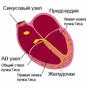 Excitement spreading through conducting system, sequentially covers the parts of the heart. Starting in the sinoatrial (sinus) node (the wall of the right atrium), which has maximum automaticity, the impulse passes through the atrial muscles, the atrioventricular node, the bundle of His with its legs and is directed to the ventricles, stimulating parts of the conduction system even before the manifestation of its own automaticity .
Excitation that occurs on the outer surface of the myocardium leaves this part electronegative in relation to areas not affected by excitation. However, due to the fact that body tissues have electrical conductivity, biocurrents are projected onto the surface of the body and can be recorded and recorded on a moving tape in the form of a curve - an electrocardiogram. The ECG consists of waves that are repeated after each heartbeat, and through them shows the disorders that exist in the human heart.
How is an ECG taken?
Many people can probably answer this question. Doing an ECG, if necessary, will also not be difficult - there is an electrocardiograph in every clinic. ECG technique? It only seems at first glance that it is so familiar to everyone, but meanwhile, only medical workers who have undergone special training in taking an electrocardiogram know it. But we hardly need to go into details, since no one will allow us to do such work without preparation anyway.

Patients need to know how to properly prepare: that is, it is advisable not to overeat, not to smoke, not to drink alcoholic beverages and medications, not to get involved in heavy physical labor and not to drink coffee before the procedure, otherwise you can fool the ECG. It will certainly be provided, if nothing else.
So, a completely calm patient undresses to the waist, frees his legs and lies down on the couch, and the nurse will lubricate the necessary places (leads) with a special solution, apply electrodes from which wires of different colors go to the device, and take a cardiogram.
The doctor will decipher it later, but if you are interested, you can try to figure out your teeth and intervals yourself.
Teeth, leads, intervals
This section may not be of interest to everyone, in which case you can skip it, but for those who are trying to understand their ECG on their own, it may be useful.
The waves in the ECG are designated using Latin letters: P, Q, R, S, T, U, where each of them reflects the state of different parts of the heart:
- P – atrial depolarization;
- QRS wave complex – ventricular depolarization;
- T – ventricular repolarization;
- A weak U wave may indicate repolarization of the distal portions of the ventricular conduction system.

To record an ECG, 12 leads are usually used:
- 3 standard – I, II, III;
- 3 reinforced unipolar limb leads (according to Goldberger);
- 6 reinforced unipolar chest (according to Wilson).
In some cases (arrhythmias, abnormal location of the heart), there is a need to use additional unipolar chest and bipolar leads according to Neb (D, A, I).

When interpreting the ECG results, the duration of the intervals between its components is measured. This calculation is necessary to assess the rhythm frequency, where the shape and size of the teeth in different leads will be an indicator of the nature of the rhythm, the electrical phenomena occurring in the heart and (to some extent) the electrical activity of individual sections of the myocardium, that is, the electrocardiogram shows how our heart works at that time. or another period.
Video: lesson on ECG waves, segments and intervals
ECG analysis
A more rigorous interpretation of the ECG is made by analyzing and calculating the area of the teeth when using special leads (vector theory), however, in practice, they mainly make do with such an indicator as electrical axis direction, which is the total QRS vector. It is clear that everyone’s chest is structured differently and the heart does not have such a strict arrangement, the weight ratio of the ventricles and the conductivity inside them are also different for everyone, therefore, when deciphering, the horizontal or vertical direction of this vector is indicated.
Doctors carry out ECG analysis in a sequential order, determining the norm and violations:
- Assess the heart rhythm and measure the heart rate (with a normal ECG - sinus rhythm, heart rate - from 60 to 80 beats per minute);
- Intervals (QT, norm – 390-450 ms) are calculated, characterizing the duration of the contraction phase (systole) using a special formula (I often use Bazett’s formula). If this interval lengthens, the doctor has the right to suspect. Hypercalcemia, on the contrary, leads to a shortening of the QT interval. The conductivity of the pulses reflected through the intervals is calculated using a computer program, which significantly increases the reliability of the results;
- they begin to calculate from the isoline according to the height of the teeth (normally R is always higher than S) and if S exceeds R and the axis deviates to the right, then they think about disturbances in the activity of the right ventricle, if on the contrary - to the left, and the height of S is greater than R in II and III leads – left ventricular hypertrophy is suspected;
- The QRS complex is studied, which is formed during the conduction of electrical impulses to the ventricular muscle and determines the activity of the latter (the norm is the absence of a pathological Q wave, the width of the complex is not more than 120 ms). If this interval shifts, then we speak of blockades (full or partial) of the bundle branches or conduction disturbances. Moreover, incomplete blockade of the right bundle branch is an electrocardiographic criterion of right ventricular hypertrophy, and incomplete blockade of the left bundle branch may indicate left ventricular hypertrophy;
- They describe the ST segments, which reflect the period of restoration of the initial state of the heart muscle after its complete depolarization (normally located on the isoline) and the T wave, which characterizes the process of repolarization of both ventricles, which is directed upward, asymmetrical, its amplitude is lower than the wave in duration and is longer than the QRS complex.
The decoding work is carried out only by a doctor, however, some ambulance paramedics perfectly recognize common pathologies, which is very important in emergency cases. But first, you still need to know the ECG norm.

This is what the cardiogram of a healthy person looks like, whose heart works rhythmically and correctly, but not everyone knows what this record means, which can change under various physiological conditions, such as pregnancy. In pregnant women, the heart takes a different position in the chest, so the electrical axis shifts. In addition, depending on the duration, the load on the heart is added. An ECG during pregnancy will reflect these changes.
The cardiogram indicators in children are also excellent; they will “grow” with the baby, and therefore will change according to age; only after 12 years, the child’s electrocardiogram begins to approach the ECG of an adult.
The most disappointing diagnosis: heart attack

The most serious diagnosis on the ECG, of course, is, in the recognition of which the cardiogram plays the main role, because it is she (the first!) that finds areas of necrosis, determines the localization and depth of the lesion, and can distinguish an acute infarction from the scars of the past.
The classic signs of myocardial infarction on the ECG are the registration of a deep Q wave (OS), segment elevationST, which deforms R, smoothing it, and the subsequent appearance of a negative pointed isosceles tooth T. This elevation of the ST segment visually resembles a cat’s back (“cat”). However, a distinction is made between myocardial infarction with and without the Q wave.

Video: signs of a heart attack on an ECG
When there's something wrong with your heart
Often in ECG conclusions you can find the expression: “”. As a rule, such a cardiogram is obtained by people whose hearts have had an additional load for a long time, for example, due to obesity. It is clear that the left ventricle has a hard time in such situations. Then the electrical axis deviates to the left, and S becomes greater than R.

hypertrophy of the left (left) and right (right) ventricles of the heart on the ECG
Video: cardiac hypertrophy on ECG
One of the presenters will answer your question.
The questions in this section are currently answered by: Sazykina Oksana Yurievna, cardiologist, therapist
You can thank a specialist for their help or support the VesselInfo project at any time.
In questions about interpreting the ECG, be sure to indicate the patient’s gender, age, clinical data, diagnoses and complaints.
In this issue I will briefly touch on these issues.
Also, previous issues and materials for a more in-depth study of ECG can be found in the "" section.
Numerous ECG manuals describe a fairly large number of electrocardiographic signs hypertrophy myocardium.
Thus, M. S. Kushakovsky (1986) points to 136 signs of myocardial hypertrophy that can be determined on an ECG.
1. What signs of myocardial hypertrophy are there?
1. In hypertrophied myocardium, excitation will take much longer to travel from the endocardium to the epicardium than in normal myocardium.
Increasing internal deviation time - first ECG sign of hypertrophy
2. In the hypertrophied myocardium, the excitation vector going from the endocardium to the epicardium is larger in magnitude compared to the norm.
Consequently, a recording electrode located above the hypertrophied myocardium will graphically display this vector on the ECG as a K wave much larger in amplitude than the normal R wave.
Increase in R wave amplitude - second ECG sign of hypertrophy.
3. Blood supply to the myocardium is carried out through the coronary arteries, which are located subepicardially. In myocardium of normal thickness, the subendocardial layers are adequately supplied with blood. As the thickness of the myocardium increases, the subendocardial layers begin to experience a lack (deficiency) of blood flowing through the coronary arteries. Deficiency or lack of blood is ischemia - ischemic (lat.).
Ischemia of the subendocardial layers of the myocardium - third ECG sign of hypertrophy.
4. The ventricular conduction system is anatomically located under the endocardium. With ischemia of the subendocardial layers of the myocardium, the function of the conduction pathways will be impaired to a certain extent.
Conduction disturbances in hypertrophied myocardium - fourth ECG sign of hypertrophy.
5. In the case of hypertrophy of one of the ventricles, its mass increases due to the growth of cardiomyocytes. Its excitation vector will become greater than the excitation vector of the non-hypertrophied ventricle, and the resulting vector will deviate towards the hypertrophied ventricle. The electrical axis of the heart is inextricably linked with the resulting vector, which during hypertrophy will deviate from its normal position.
Deviation of the electrical axis of the heart towards the hypertrophied ventricle - fifth ECG sign of hypertrophy.
6. The electrical position of the heart is also inextricably linked with the direction of the resulting vector. When the direction of the resulting vector changes, due to hypertrophy, the electrical position of the heart will change.
Change in the electrical position of the heart - sixth ECG sign of hypertrophy.
7. With a normal position of the electrical axis of the heart and the basic electrical position of the heart, the third chest lead (V3) is a transition zone.
The transition zone is called such a chest lead in which the height of the R wave and the depth of the S wave are equal in absolute value. Naturally, when the electrical axis and electrical position of the heart changes, the ratio of the R and S waves in the third chest lead will change. The transition zone will shift to another chest lead (to the lead where the equality of the values of the R and S waves will remain).
Transition zone offset - the seventh ECG is a sign of hypertrophy.
2. What are the signs of left ventricular myocardial hypertrophy?

1. An increase in the time of internal deviation in the left chest leads V5 and V6 by more than 0.05 s.
2. An increase in the amplitude of the K wave in the left leads - I, aVL, V5 and V6.
3. Displacement of the S-T segment below the isoelectric line, inversion or biphasicity of the T wave in the left leads - I, aVL, V5 and Vb.
4. Conduction disturbance along the left bundle branch: complete or incomplete blockade of the bundle.
5. Deviation of the electrical axis of the heart to the left (left-gram)
6. Horizontal or semi-horizontal electrical position of the heart.
7. Displacement of the transition zone into lead V2 or V1.
3. What are the signs of right ventricular myocardial hypertrophy?

1. An increase in the time of internal deviation in the right chest leads V1 and V2 by more than 0.03 s.
2. Increase in the amplitude of the K wave in the right leads III, aVF, V1 and V2.
3. Displacement of the S-T segment below the isoelectric line, inversion or biphasicity of the T wave in the right leads - I I I, aVF, V1 and V2.
4. Impaired conduction along the right bundle branch: complete or incomplete bundle block.
5. Deviation of the electrical axis of the heart to the right (right-gram).
6. Vertical or semi-vertical electrical position of the heart.
7. Shift of the transition zone to lead V4 or V5.
4. What are the signs of atrial hypertrophy?
The P wave represents the summative excitation of both atria. In the case of right atrium hypertrophy, the width and height of its excitation peak will increase (1st and 2nd electrocardiographic sign of hypertrophy). This condition will lead to the fact that the summation peak of atrial excitation - the P wave - will become higher in amplitude.
  In some cases, its outline takes on a pointed shape in the form of a tent. Since hypertrophy of the right atrium is observed more often in diseases of the lungs, the modified P wave in these cases is also called P-pulmonale. With hypertrophy of the left atrium, the width and height of the peak, reflecting its excitation, increase.
  The summation wave P will become wide, its outline taking on the shape of a double hump. Most often, left atrial hypertrophy is observed with mitral heart defects. Therefore, the P wave with left atrial hypertrophy is called P-mitrale. Thus, electrocardiographic signs of atrial hypertrophy are: right atrium - increased amplitude and sharpness of the P wave; it is often called P-pulmonale; left atrium - widening of the P wave by more than 0.12 s and its double hump; such a tooth is called P-mitrale.
 Conclusions from this newsletter:
1. There are a number of additional methods that allow you to accurately determine myocardial hypertrophy. These include cardiac ultrasound, nuclear magnetic resonance, computed x-ray tomography, and x-ray diagnostics. Electrocardiography does not accurately detect anatomical myocardial hypertrophy. However, it is useful to know the ECG signs of hypertrophy both for further assimilation of the material and for understanding a number of clinical situations.
2. There are many electrocardiographic signs of hypertrophy.
3. Of the many of these signs, we have identified the 7 most important in the diagnosis of ventricular hypertrophy.
4. It is not at all necessary to have all the signs of hypertrophy on the ECG at once. In some cases, only a few of them can be identified.
5. The first and second signs are associated with the passage of a single vector through the myocardium from the endocardium to the epicardium.
6. The third and fourth signs characterize myocardial hypertrophy with overload.
7. The fifth, sixth and seventh signs are caused by a change in the resulting vector of ventricular excitation.
Conclusion.
Even more information to explore ECG in the form of articles and video lessons In chapter " ".
Sincerely yours website.Addition to the newsletter release "Electrocardiographic signs of myocardial hypertrophy":
The P wave in the form of P-mitrale is indeed observed with hypertrophy of the left atrium. However, the P wave, exactly the same in width (more than 0.12 s) and in shape (double hump), is recorded on the electrocardiogram when intraatrial conduction is disrupted, otherwise called intraatrial block. You, of course, noticed that one of the ECG signs of myocardial hypertrophy is conduction disturbances. Finally, the electrical axis of the heart, significantly deviating during hypertrophy to the left (alpha angle less than - 30°) or to the right (alpha angle greater than +90°), indicates blockade of the branches of the left bundle branch.
In other words, electrocardiographic signs of hypertrophy are closely related to electrocardiographic signs of conduction disturbances, to which we now proceed.
Educational video ECG for atrial and ventricular hypertrophy
If you have problems watching, download the video from the pageThank you
The site provides reference information for informational purposes only. Diagnosis and treatment of diseases must be carried out under the supervision of a specialist. All drugs have contraindications. Consultation with a specialist is required!
Electrocardiogram is a widely used method of objective diagnostics various pathologies of the human heart, which is used almost everywhere today. An electrocardiogram (ECG) is taken in a clinic, in an ambulance, or in a hospital department. ECG is a very important recording that reflects the condition of the heart. That is why the reflection of various types of cardiac pathology on the ECG is described by a separate science - electrocardiography. Electrocardiography also deals with the problems of correct ECG recording, decoding issues, interpretation of controversial and unclear points, etc.Definition and essence of the method
An electrocardiogram is a recording of the heart, which is presented as a curved line on paper. The cardiogram line itself is not chaotic; it has certain intervals, teeth and segments that correspond to certain stages of the heart.To understand the essence of an electrocardiogram, you need to know what exactly is recorded by a device called an electrocardiograph. The ECG records the electrical activity of the heart, which changes cyclically in accordance with the onset of diastole and systole. The electrical activity of the human heart may seem like fiction, but this unique biological phenomenon exists in reality. In reality, the heart contains so-called cells of the conduction system, which generate electrical impulses that are transmitted to the muscles of the organ. It is these electrical impulses that cause the myocardium to contract and relax with a certain rhythm and frequency.
The electrical impulse propagates through the cells of the conduction system of the heart strictly sequentially, causing contraction and relaxation of the corresponding sections - the ventricles and atria. The electrocardiogram reflects precisely the total electrical potential difference in the heart.
decryption?
 An electrocardiogram can be taken in any clinic or multidisciplinary hospital. You can contact a private medical center where there is a specialist cardiologist or therapist. After recording the cardiogram, the tape with curves is examined by the doctor. It is he who analyzes the recording, deciphers it and writes a final report, which reflects all visible pathologies and functional deviations from the norm.
 An electrocardiogram is recorded using a special device - an electrocardiograph, which can be multi-channel or single-channel. The speed of ECG recording depends on the modification and modernity of the device. Modern devices can be connected to a computer, which, with a special program, will analyze the recording and issue a final conclusion immediately after the procedure is completed.
Any cardiograph has special electrodes that are applied in a strictly defined order. There are four clothespins in red, yellow, green and black that are placed on both arms and both legs. If you go in a circle, then the clothespins are applied according to the rule “red-yellow-green-black”, from the right hand. It's easy to remember this sequence thanks to the student saying: "Every-Woman is an Eviler Trait." In addition to these electrodes, there are also chest electrodes, which are installed in the intercostal spaces.
As a result, the electrocardiogram consists of twelve waveforms, six of which are recorded from the chest electrodes, and are called chest leads. The remaining six leads are recorded from electrodes attached to the arms and legs, with three of them called standard and three more called enhanced. The chest leads are designated V1, V2, V3, V4, V5, V6, the standard ones are simply Roman numerals - I, II, III, and the reinforced leg leads - the letters aVL, aVR, aVF. Different leads of the cardiogram are necessary to create the most complete picture of the activity of the heart, since some pathologies are visible on the chest leads, others on the standard ones, and still others on the enhanced ones.
The person lies down on the couch, the doctor attaches the electrodes and turns on the device. While the ECG is being written, the person must be absolutely calm. We must not allow the appearance of any irritants that can distort the true picture of the work of the heart.
How to correctly perform an electrocardiogram followed by
transcript - video
The principle of deciphering an ECG
Since the electrocardiogram reflects the processes of contraction and relaxation of the myocardium, it is possible to trace how these processes proceed and identify existing pathological processes. The elements of the electrocardiogram are closely related and reflect the duration of the phases of the cardiac cycle - systole and diastole, that is, contraction and subsequent relaxation. Decoding the electrocardiogram is based on the study of the teeth, their position relative to each other, duration, and other parameters. The following elements of the electrocardiogram are studied for analysis:1. Teeth.
2. Intervals.
3. Segments.
All sharp and smooth convexities and concavities on the ECG line are called teeth. Each tooth is designated by a letter of the Latin alphabet. The P wave reflects contraction of the atria, the QRS complex – contraction of the ventricles of the heart, the T wave – relaxation of the ventricles. Sometimes after the T wave on the electrocardiogram there is another U wave, but it has no clinical and diagnostic role.
An ECG segment is considered to be a segment enclosed between adjacent teeth. For diagnosing heart pathology, the P – Q and S – T segments are of great importance. The interval on the electrocardiogram is a complex that includes a tooth and an interval. The P–Q and Q–T intervals are of great importance for diagnosis.
Often in the doctor’s report you can see small Latin letters, which also indicate teeth, intervals and segments. Small letters are used if the prong is less than 5 mm long. In addition, several R waves may appear in the QRS complex, which are usually designated R’, R”, etc. Sometimes the R wave is simply missing. Then the entire complex is designated by only two letters - QS. All this has important diagnostic value.
ECG interpretation plan - general scheme for reading results
 When deciphering an electrocardiogram, the following parameters reflecting the work of the heart must be established:
 - position of the electrical axis of the heart;
- determining the correctness of the heart rhythm and conductivity of the electrical impulse (blockades, arrhythmias are identified);
- determining the regularity of contractions of the heart muscle;
- determination of heart rate;
- identifying the source of the electrical impulse (whether sinus rhythm is determined or not);
- analysis of the duration, depth and width of the atrial P wave and the P – Q interval;
- analysis of the duration, depth, width of the QRST ventricular wave complex;
- analysis of parameters of the RS – T segment and T wave;
- analysis of Q – T interval parameters.
In the conclusion on the electrocardiogram, the doctor must reflect the following parameters:
- sinus rhythm or not;
- rhythm regularity;
- heart rate (HR);
- position of the electrical axis of the heart.
Example of deciphering an electrocardiogram
At the very beginning of the electrocardiogram tape there should be a calibration signal, which looks like a large letter “P” 10 mm high. If this calibration signal is not present, then the electrocardiogram is uninformative. If the height of the calibration signal is below 5 mm in standard and enhanced leads, and below 8 mm in chest leads, then there is a low voltage of the electrocardiogram, which is a sign of a number of heart pathologies. For subsequent decoding and calculation of some parameters, you need to know what period of time fits into one cell of graph paper. At a belt speed of 25 mm/s, one cell 1 mm long is equal to 0.04 seconds, and at a speed of 50 mm/s – 0.02 seconds.Checking the regularity of heart contractions
It is assessed by the intervals R - R. If the teeth are located at the same distance from each other throughout the entire recording, then the rhythm is regular. Otherwise it is called correct. Estimating the distance between the R - R teeth is very simple: the electrocardiogram is recorded on graph paper, which makes it easy to measure any gaps in millimeters.Heart rate (HR) calculation
 It is carried out using a simple arithmetic method: count the number of large squares on graph paper that are placed between two R waves. Then the heart rate is calculated using the formula, which is determined by the speed of the tape in the cardiograph:
 1. The tape speed is 50 mm/s - then the heart rate is 600 divided by the number of squares.
2. The tape speed is 25 mm/s - then the heart rate is 300 divided by the number of squares.
For example, if 4.8 large squares fit between two R teeth, then the heart rate, at a belt speed of 50 mm/s, will be equal to 600/4.8 = 125 beats per minute.
If the heart rate is abnormal, then the maximum and minimum heart rate is determined, also taking as a basis the maximum and minimum distances between the R waves.
Identifying the source of the rhythm
The doctor studies the rhythm of heart contractions and finds out which node of nerve cells causes the cyclic processes of contraction and relaxation of the heart muscle. This is very important for identifying blockages.Decoding ECG - rhythms
Normally, the pacemaker is the sinus node. And such a normal rhythm itself is called sinus - all other options are pathological. In various pathologies, any other node of the nerve cells of the cardiac conduction system can act as a pacemaker. In this case, the cyclic electrical impulses become confused and the heart rhythm is disrupted - an arrhythmia occurs.In sinus rhythm on the electrocardiogram in lead II there is a P wave before each QRS complex, and it is always positive. In one lead, all P waves should have the same shape, length and width.
With atrial rhythm the P wave in leads II and III is negative, but is present before each QRS complex.
Atrioventricular rhythms are characterized by the absence of P waves on cardiograms, or the appearance of this wave after the QRS complex, and not before it, as is normal. With this type of rhythm, the heart rate is low, ranging from 40 to 60 beats per minute.
Ventricular rhythm characterized by an increase in the width of the QRS complex, which becomes large and quite frightening. The P waves and the QRS complex are completely unrelated to each other. That is, there is no strict correct normal sequence - the P wave, followed by the QRS complex. Ventricular rhythm is characterized by a decrease in heart rate - less than 40 beats per minute.
Detection of pathology of electrical impulse conduction through the structures of the heart
To do this, measure the duration of the P wave, the P–Q interval and the QRS complex. The duration of these parameters is calculated from the millimeter tape on which the cardiogram is recorded. First, count how many millimeters each tooth or interval occupies, after which the resulting value is multiplied by 0.02 at a recording speed of 50 mm/s, or by 0.04 at a recording speed of 25 mm/s.The normal duration of the P wave is up to 0.1 seconds, the P – Q interval is 0.12-0.2 seconds, the QRS complex is 0.06-0.1 seconds.
Electrical axis of the heart
 Denoted as the alpha angle. It can have a normal position, horizontal or vertical. Moreover, in a thin person the axis of the heart is more vertical relative to the average values, while in a fat person it is more horizontal. The normal position of the electrical axis of the heart is 30–69 o, vertical – 70–90 o, horizontal – 0–29 o. The alpha angle, equal to 91 to ±180 o, reflects a sharp deviation of the electrical axis of the heart to the right. The alpha angle, equal to 0 to –90 o, reflects a sharp deviation of the electrical axis of the heart to the left.
 The electrical axis of the heart can deviate under various pathological conditions. For example, hypertension leads to a deviation to the right; a conduction disorder (blockade) can shift it to the right or left.
Atrial P wave
The atrial P wave should be:- positive in I, II, aVF and chest leads (2, 3,4, 5, 6);
- negative in aVR;
- biphasic (part of the tooth lies in the positive region, and part in the negative) in III, aVL, V1.
Pathological forms of the P wave may indicate the following pathologies:
1.
Tall and sharp teeth in leads II, III, aVF appear with hypertrophy of the right atrium (“cor pulmonale”);
2.
A P wave with two peaks and a large width in leads I, aVL, V5 and V6 indicates hypertrophy of the left atrium (for example, mitral valve disease).
P–Q interval
The P–Q interval has a normal duration of 0.12 to 0.2 seconds. An increase in the duration of the P–Q interval is a reflection of atrioventricular block. On the electrocardiogram, three degrees of atrioventricular block (AV) can be distinguished:- I degree: simple lengthening of the P–Q interval while preserving all other complexes and waves.
- II degree: prolongation of the P–Q interval with partial loss of some QRS complexes.
- III degree: lack of connection between the P wave and QRS complexes. In this case, the atria work in their own rhythm, and the ventricles - in their own.
Ventricular QRST complex
 The ventricular QRST complex consists of the QRS complex itself and the S – T segment. The normal duration of the QRST complex does not exceed 0.1 seconds, and its increase is detected with blockades of the Hiss bundle branches.
 QRS complex consists of three waves, Q, R and S, respectively. The Q wave is visible on the cardiogram in all leads except 1, 2 and 3 chest leads. A normal Q wave has an amplitude up to 25% of that of an R wave. The duration of the Q wave is 0.03 seconds. The R wave is recorded in absolutely all leads. The S wave is also visible in all leads, but its amplitude decreases from the 1st thoracic to the 4th, and in the 5th and 6th it may be completely absent. The maximum amplitude of this tooth is 20 mm.
The S–T segment is very important from a diagnostic point of view. It is by this tooth that myocardial ischemia can be detected, that is, a lack of oxygen in the heart muscle. Usually this segment runs along the isoline, in the 1st, 2nd and 3rd chest leads; it can rise up by a maximum of 2 mm. And in the 4th, 5th and 6th chest leads, the S-T segment can shift below the isoline by a maximum of half a millimeter. It is the deviation of the segment from the isoline that reflects the presence of myocardial ischemia.
T wave
The T wave is a reflection of the process of eventual relaxation in the cardiac muscle of the ventricles of the heart. Typically, when the amplitude of the R wave is large, the T wave will also be positive. A negative T wave is normally recorded only in lead aVR.Q-T interval
The Q–T interval reflects the process of eventual contraction in the myocardium of the ventricles of the heart.ECG interpretation - normal indicators
The transcript of the electrocardiogram is usually recorded by the doctor in conclusion. A typical example of a normal cardiac cardiogram looks like this:1. PQ – 0.12 s.
2. QRS – 0.06 s.
3. QT – 0.31 s.
4. RR – 0.62 – 0.66 – 0.6.
5. Heart rate is 70 - 75 beats per minute.
6. sinus rhythm.
7. The electrical axis of the heart is located normally.
Normally, the rhythm should be only sinus, the heart rate of an adult is 60 - 90 beats per minute. The P wave is normally no more than 0.1 s, the P – Q interval is 0.12-0.2 seconds, the QRS complex is 0.06-0.1 seconds, Q – T is up to 0.4 s.
If the cardiogram is pathological, then it indicates specific syndromes and deviations from the norm (for example, partial blockade of the left bundle branch, myocardial ischemia, etc.). The doctor can also reflect specific violations and changes in the normal parameters of the waves, intervals and segments (for example, shortening of the P wave or Q-T interval, etc.).
Interpretation of ECG in children and pregnant women
In principle, children and pregnant women have normal heart electrocardiogram readings - the same as in healthy adults. However, there are certain physiological characteristics. For example, the heart rate of children is higher than that of an adult. The normal heart rate of a child up to 3 years of age is 100–110 beats per minute, 3–5 years old – 90–100 beats per minute. Then gradually the heart rate decreases, and in adolescence it is compared with that of an adult - 60 - 90 beats per minute.In pregnant women, there may be a slight deviation of the electrical axis of the heart in late gestation due to compression by the growing uterus. In addition, sinus tachycardia often develops, that is, an increase in heart rate to 110 - 120 beats per minute, which is a functional condition and goes away on its own. An increase in heart rate is associated with a greater volume of circulating blood and increased workload. Due to the increased load on the heart, pregnant women may experience overload in various parts of the organ. These phenomena are not a pathology - they are associated with pregnancy and will go away on their own after childbirth.
Decoding the electrocardiogram during a heart attack
 Myocardial infarction is a sudden cessation of oxygen supply to the heart muscle cells, resulting in the development of necrosis of a tissue area that is in a state of hypoxia. The reason for the disruption of oxygen supply can be different - most often it is a blockage of a blood vessel, or its rupture. A heart attack involves only part of the muscle tissue of the heart, and the extent of the damage depends on the size of the blood vessel that is blocked or ruptured. On an electrocardiogram, myocardial infarction has certain signs by which it can be diagnosed.
 In the process of development of myocardial infarction, four stages are distinguished, which have different manifestations on the ECG:
- acute;
- acute;
- subacute;
- cicatricial.
Sometimes it is possible to detect the phase of myocardial ischemia preceding the acute phase, which is characterized by high T waves.
Acute stage A heart attack lasts 2–3 weeks. During this period, a wide and high-amplitude Q wave and a negative T wave are recorded on the ECG.
Subacute stage lasts up to 3 months. The ECG shows a very large negative T wave with a huge amplitude, which gradually normalizes. Sometimes a rise in the S-T segment is detected, which should have leveled off by this period. This is an alarming symptom, as it may indicate the formation of a cardiac aneurysm.
Scar stage heart attack is final, since connective tissue is formed at the damaged site, incapable of contraction. This scar is recorded on the ECG as a Q wave, which will remain for life. Often the T wave is smoothed, has a low amplitude, or is completely negative.
Interpretation of the most common ECGs
 In conclusion, doctors write the result of the ECG interpretation, which is often incomprehensible because it consists of terms, syndromes and simply statements of pathophysiological processes. Let's consider the most common ECG conclusions, which are incomprehensible to a person without a medical education.
 Ectopic rhythm means not sinus - which can be either a pathology or a norm. The norm is ectopic rhythm when there is a congenital malformation of the conduction system of the heart, but the person does not present any complaints and does not suffer from other cardiac pathologies. In other cases, an ectopic rhythm indicates the presence of blockades.
Changes in repolarization processes on the ECG reflects a violation of the process of relaxation of the heart muscle after contraction.
Sinus rhythm This is the normal heart rate of a healthy person.
Sinus or sinusoidal tachycardia means that a person has a correct and regular rhythm, but an increased heart rate - more than 90 beats per minute. In young people under 30 years of age, this is a variant of the norm.
Sinus bradycardia- this is a low heart rate - less than 60 beats per minute against the background of a normal, regular rhythm.
Nonspecific ST-T changes mean that there are minor deviations from the norm, but their cause may be completely unrelated to heart pathology. It is necessary to undergo a full examination. Such nonspecific ST-T changes can develop with an imbalance of potassium, sodium, chlorine, magnesium ions, or various endocrine disorders, often during menopause in women.
Biphasic R wave in combination with other signs of a heart attack indicates damage to the anterior wall of the myocardium. If no other signs of a heart attack are detected, then a biphasic R wave is not a sign of pathology.
QT prolongation may indicate hypoxia (lack of oxygen), rickets, or overexcitation of the child’s nervous system, which is a consequence of birth trauma.
Myocardial hypertrophy means that the muscular wall of the heart is thickened and works under enormous load. This can lead to the formation of:
- heart failure;
- arrhythmias.
Moderate diffuse changes in the myocardium mean that tissue nutrition is impaired and cardiac muscle dystrophy has developed. This is a fixable condition: you need to see a doctor and undergo an adequate course of treatment, including normalizing your diet.
Deviation of the electrical axis of the heart (EOS) left or right is possible with hypertrophy of the left or right ventricle, respectively. EOS can deviate to the left in obese people, and to the right - in thin people, but in this case this is a variant of the norm.
Left type ECG– EOS deviation to the left.
NBPNPG– an abbreviation for “incomplete right bundle branch block.” This condition can occur in newborns and is a normal variant. In rare cases, RBBB can cause arrhythmia, but generally does not lead to the development of negative consequences. Block of the Hiss bundle branch is quite common in people, but if there are no complaints about the heart, then it is not at all dangerous.
BPVLNPG– an abbreviation meaning “blockade of the anterior branch of the left bundle branch.” Reflects a violation of the conduction of electrical impulses in the heart, and leads to the development of arrhythmias.
Small growth of the R wave in V1-V3 may be a sign of interventricular septal infarction. To accurately determine whether this is the case, it is necessary to do another ECG study.
CLC syndrome(Klein-Levy-Kritesco syndrome) is a congenital feature of the conduction system of the heart. May cause the development of arrhythmias. This syndrome does not require treatment, but it is necessary to be regularly examined by a cardiologist.
Low voltage ECG often recorded with pericarditis (a large amount of connective tissue in the heart that has replaced muscle tissue). In addition, this sign may be a reflection of exhaustion or myxedema.
Metabolic changes are a reflection of insufficient nutrition of the heart muscle. It is necessary to be examined by a cardiologist and undergo a course of treatment.
Conduction slowdown means that the nerve impulse travels through the tissues of the heart more slowly than normal. This condition itself does not require special treatment - it may be a congenital feature of the conduction system of the heart. Regular monitoring by a cardiologist is recommended.
Blockade 2 and 3 degrees reflects a serious disturbance of cardiac conduction, which is manifested by arrhythmia. In this case, treatment is necessary.
Rotation of the heart by the right ventricle forward may be an indirect sign of the development of hypertrophy. In this case, it is necessary to find out its cause and undergo a course of treatment, or adjust your diet and lifestyle.
Price of an electrocardiogram with interpretation
The cost of an electrocardiogram with interpretation varies significantly, depending on the specific medical institution. Thus, in public hospitals and clinics the minimum price for the procedure of taking an ECG and interpreting it by a doctor is from 300 rubles. In this case, you will receive films with recorded curves and a doctor’s conclusion on them, which he will make himself, or using a computer program.If you want to receive a thorough and detailed conclusion on the electrocardiogram, a doctor’s explanation of all the parameters and changes, it is better to contact a private clinic that provides similar services. Here the doctor will be able not only to write a conclusion by deciphering the cardiogram, but also to calmly talk to you, taking his time to explain all the points of interest. However, the cost of such a cardiogram with interpretation in a private medical center ranges from 800 rubles to 3,600 rubles. You should not assume that bad specialists work in an ordinary clinic or hospital - it’s just that a doctor in a public institution, as a rule, has a very large amount of work, so he simply does not have time to talk with each patient in great detail.
Decoding an ECG is the job of a knowledgeable doctor. This method of functional diagnostics evaluates:
- heart rate - the state of the generators of electrical impulses and the state of the heart system conducting these impulses
- condition of the heart muscle itself (myocardium), the presence or absence of inflammation, damage, thickening, oxygen starvation, electrolyte imbalance
However, modern patients often have access to their medical documents, in particular, to electrocardiography films on which medical reports are written. With their diversity, these records can reach even the most balanced but ignorant person. After all, the patient often does not know for certain how dangerous to life and health is what is written on the back of the ECG film by the hand of a functional diagnostician, and there are still several days before an appointment with a therapist or cardiologist.
To reduce the intensity of passions, we immediately warn readers that with not a single serious diagnosis (myocardial infarction, acute rhythm disturbances), a functional diagnostician will not let a patient leave the office, but, at a minimum, will send him for a consultation with a fellow specialist right there. About the rest of the “open secrets” in this article. In all unclear cases of pathological changes in the ECG, ECG monitoring, 24-hour monitoring (Holter), ECHO cardioscopy (ultrasound of the heart) and stress tests (treadmill, bicycle ergometry) are prescribed.
Numbers and Latin letters in ECG interpretation
PQ- (0.12-0.2 s) – atrioventricular conduction time. Most often it lengthens against the background of AV blockade. Shortened in CLC and WPW syndromes.
P – (0.1s) height 0.25-2.5 mm describes atrial contractions. May indicate their hypertrophy.
QRS – (0.06-0.1s) -ventricular complex
QT – (no more than 0.45 s) lengthens with oxygen starvation (myocardial ischemia, infarction) and the threat of rhythm disturbances.
RR - the distance between the apices of the ventricular complexes reflects the regularity of heart contractions and makes it possible to calculate heart rate.
The interpretation of the ECG in children is presented in Fig. 3
Heart Rate Description Options
Sinus rhythm
This is the most common inscription found on an ECG. And, if nothing else is added and the frequency (HR) is indicated from 60 to 90 beats per minute (for example, HR 68`) - this is the best option, indicating that the heart works like a clock. This is the rhythm set by the sinus node (the main pacemaker that generates electrical impulses that cause the heart to contract). At the same time, sinus rhythm implies well-being, both in the state of this node and the health of the conduction system of the heart. The absence of other records denies pathological changes in the heart muscle and means that the ECG is normal. In addition to sinus rhythm, there may be atrial, atrioventricular or ventricular, indicating that the rhythm is set by cells in these parts of the heart and is considered pathological.
Sinus arrhythmia
This is a normal variant in young people and children. This is a rhythm in which impulses leave the sinus node, but the intervals between heart contractions are different. This may be due to physiological changes (respiratory arrhythmia, when heart contractions slow down during exhalation). Approximately 30% of sinus arrhythmias require observation by a cardiologist, as they are at risk of developing more serious rhythm disturbances. These are arrhythmias after rheumatic fever. Against the background of myocarditis or after it, against the background of infectious diseases, heart defects and in persons with a family history of arrhythmias.
Sinus bradycardia
These are rhythmic contractions of the heart with a frequency of less than 50 per minute. In healthy people, bradycardia occurs, for example, during sleep. Bradycardia also often occurs in professional athletes. Pathological bradycardia may indicate sick sinus syndrome. In this case, bradycardia is more pronounced (heart rate from 45 to 35 beats per minute on average) and is observed at any time of the day. When bradycardia causes pauses in heart contractions of up to 3 seconds during the day and about 5 seconds at night, leads to disturbances in the supply of oxygen to tissues and is manifested, for example, by fainting, an operation is indicated to install a cardiac pacemaker, which replaces the sinus node, imposing a normal rhythm of contractions on the heart. 
Sinus tachycardia
Heart rate more than 90 per minute is divided into physiological and pathological. In healthy people, sinus tachycardia is accompanied by physical and emotional stress, drinking coffee, sometimes strong tea or alcohol (especially energy drinks). It is short-lived and after an episode of tachycardia, the heart rate returns to normal within a short period of time after stopping the load. With pathological tachycardia, heartbeats bother the patient at rest. Its causes include fever, infections, blood loss, dehydration, anemia,. The underlying disease is treated. Sinus tachycardia is stopped only in case of a heart attack or acute coronary syndrome.
Extarsystole
These are rhythm disturbances in which foci outside the sinus rhythm give extraordinary cardiac contractions, after which there is a pause of twice the length, called compensatory. In general, the patient perceives heartbeats as uneven, rapid or slow, and sometimes chaotic. The most worrying thing is the dips in heart rate. May occur in the form of tremors, tingling, feelings of fear and emptiness in the stomach.
Not all extrasystoles are dangerous to health. Most of them do not lead to significant circulatory disorders and do not threaten either life or health. They can be functional (against the background of panic attacks, cardioneurosis, hormonal imbalances), organic (with ischemic heart disease, heart defects, myocardial dystrophy or cardiopathy, myocarditis). They can also be caused by intoxication and heart surgery. Depending on the place of occurrence, extrasystoles are divided into atrial, ventricular and anthrioventricular (arising in the node at the border between the atria and ventricles).
- Single extrasystoles most often rare (less than 5 per hour). They are usually functional and do not interfere with normal blood supply.
- Paired extrasystoles two each accompany a certain number of normal contractions. Such rhythm disturbances often indicate pathology and require further examination (Holter monitoring).
- Allorhythmias are more complex types of extrasystoles. If every second contraction is an extrasystole, this is bigymenia, if every third contraction is trigymenia, every fourth is quadrigymenia.
It is customary to divide ventricular extrasystoles into five classes (according to Laun). They are assessed during daily ECG monitoring, since the readings of a regular ECG in a few minutes may not show anything.
- Class 1 - single rare extrasystoles with a frequency of up to 60 per hour, emanating from one focus (monotopic)
- 2 – frequent monotopic more than 5 per minute
- 3 – frequent polymorphic (of different shapes) polytopic (from different foci)
- 4a – paired, 4b – group (trigymenia), episodes of paroxysmal tachycardia
- 5 – early extrasystoles
The higher the class, the more serious the violations, although today even classes 3 and 4 do not always require drug treatment. In general, if there are less than 200 ventricular extrasystoles per day, they should be classified as functional and not worry about them. For more frequent cases, ECHO CS is indicated, and sometimes cardiac MRI is indicated. It is not the extrasystole that is treated, but the disease that leads to it.
Paroxysmal tachycardia
In general, a paroxysm is an attack. A paroxysmal increase in rhythm can last from several minutes to several days. In this case, the intervals between heart contractions will be the same, and the rhythm will increase over 100 per minute (on average from 120 to 250). There are supraventricular and ventricular forms of tachycardia. This pathology is based on abnormal circulation of electrical impulses in the conduction system of the heart. This pathology can be treated. Home remedies to relieve an attack:
- holding your breath
- increased forced cough
- immersing face in cold water
WPW syndrome
Wolff-Parkinson-White syndrome is a type of paroxysmal supraventricular tachycardia. Named after the authors who described it. The appearance of tachycardia is based on the presence of an additional nerve bundle between the atria and ventricles, through which a faster impulse passes than from the main pacemaker.
As a result, an extraordinary contraction of the heart muscle occurs. The syndrome requires conservative or surgical treatment (in case of ineffectiveness or intolerance of antiarrhythmic tablets, during episodes of atrial fibrillation, and with concomitant heart defects).
CLC – syndrome (Clerk-Levi-Christesco)
is similar in mechanism to WPW and is characterized by earlier excitation of the ventricles than normal due to an additional bundle along which the nerve impulse travels. The congenital syndrome is manifested by attacks of rapid heartbeat.
Atrial fibrillation
It can be in the form of an attack or a permanent form. It manifests itself in the form of atrial flutter or fibrillation.
Atrial fibrillation

Atrial fibrillation
When flickering, the heart contracts completely irregularly (the intervals between contractions of very different durations). This is explained by the fact that the rhythm is not set by the sinus node, but by other cells of the atria.
The resulting frequency is from 350 to 700 beats per minute. There is simply no full contraction of the atria; contracting muscle fibers do not effectively fill the ventricles with blood.
As a result, the heart’s output of blood deteriorates and organs and tissues suffer from oxygen starvation. Another name for atrial fibrillation is atrial fibrillation. Not all atrial contractions reach the ventricles of the heart, so the heart rate (and pulse) will be either below normal (bradysystole with a frequency of less than 60), or normal (normosystole from 60 to 90), or above normal (tachysystole more than 90 beats per minute ).
An attack of atrial fibrillation is difficult to miss.
- It usually starts with a strong beat of the heart.
- It develops as a series of completely irregular heartbeats with a high or normal frequency.
- The condition is accompanied by weakness, sweating, dizziness.
- The fear of death is very pronounced.
- There may be shortness of breath, general agitation.
- Sometimes observed.
- The attack ends with normalization of the rhythm and the urge to urinate, during which a large amount of urine is released.
To stop an attack, they use reflex methods, drugs in the form of tablets or injections, or resort to cardioversion (stimulating the heart with an electric defibrillator). If an attack of atrial fibrillation is not eliminated within two days, the risks of thrombotic complications (pulmonary embolism, stroke) increase.
With a constant form of heartbeat flicker (when the rhythm is not restored either against the background of drugs or against the background of electrical stimulation of the heart), they become a more familiar companion to patients and are felt only during tachysystole (rapid, irregular heartbeats). The main task when detecting signs of tachysystole of a permanent form of atrial fibrillation on the ECG is to slow down the rhythm to normosystole without trying to make it rhythmic.
Examples of recordings on ECG films:
- atrial fibrillation, tachysystolic variant, heart rate 160 b'.
- Atrial fibrillation, normosystolic variant, heart rate 64 b'.
Atrial fibrillation can develop in the course of coronary heart disease, against the background of thyrotoxicosis, organic heart defects, diabetes mellitus, sick sinus syndrome, and intoxication (most often with alcohol).
Atrial flutter
These are frequent (more than 200 per minute) regular contractions of the atria and equally regular, but less frequent contractions of the ventricles. In general, flutter is more common in the acute form and is better tolerated than flicker, since circulatory disorders are less pronounced. Fluttering develops when:
- organic heart diseases (cardiomyopathies, heart failure)
- after heart surgery
- against the background of obstructive pulmonary diseases
- in healthy people it almost never occurs
Clinically, flutter is manifested by rapid rhythmic heartbeat and pulse, swelling of the neck veins, shortness of breath, sweating and weakness.
Conduction disorders
Normally, having formed in the sinus node, electrical excitation travels through the conduction system, experiencing a physiological delay of a split second in the atrioventricular node. On its way, the impulse stimulates the atria and ventricles, which pump blood, to contract. If in any part of the conduction system the impulse is delayed longer than the prescribed time, then excitation to the underlying sections will come later, and, therefore, the normal pumping work of the heart muscle will be disrupted. Conduction disturbances are called blockades. They can occur as functional disorders, but more often they are the result of drug or alcohol intoxication and organic heart disease. Depending on the level at which they arise, several types are distinguished.
Sinoatrial blockade
When the exit of an impulse from the sinus node is difficult. In essence, this leads to sick sinus syndrome, slowing of contractions to severe bradycardia, impaired blood supply to the periphery, shortness of breath, weakness, dizziness and loss of consciousness. The second degree of this blockade is called Samoilov-Wenckebach syndrome.
Atrioventricular block (AV block)
This is a delay of excitation in the atrioventricular node longer than the prescribed 0.09 seconds. There are three degrees of this type of blockade. The higher the degree, the less often the ventricles contract, the more severe the circulatory disorders.
- In the first, the delay allows each atrial contraction to maintain an adequate number of ventricular contractions.
- The second degree leaves some of the atrial contractions without ventricular contractions. It is described, depending on the prolongation of the PQ interval and the loss of ventricular complexes, as Mobitz 1, 2 or 3.
- The third degree is also called complete transverse blockade. The atria and ventricles begin to contract without interconnection.
In this case, the ventricles do not stop because they obey the pacemakers from the underlying parts of the heart. If the first degree of blockade may not manifest itself in any way and can be detected only with an ECG, then the second is already characterized by sensations of periodic cardiac arrest, weakness, and fatigue. With complete blockades, brain symptoms are added to the manifestations (dizziness, spots in the eyes). Morgagni-Adams-Stokes attacks may develop (when the ventricles escape from all pacemakers) with loss of consciousness and even convulsions.
Impaired conduction within the ventricles
In the ventricles, the electrical signal propagates to the muscle cells through such elements of the conduction system as the trunk of the His bundle, its legs (left and right) and branches of the legs. Blockades can occur at any of these levels, which is also reflected in the ECG. In this case, instead of being simultaneously covered by excitation, one of the ventricles is delayed, since the signal to it bypasses the blocked area.
In addition to the place of origin, a distinction is made between complete or incomplete blockade, as well as permanent and non-permanent blockade. The causes of intraventricular blocks are similar to other conduction disorders (ischemic heart disease, myocarditis and endocarditis, cardiomyopathies, heart defects, arterial hypertension, fibrosis, heart tumors). Also affected are the use of antiarthmic drugs, an increase in potassium in the blood plasma, acidosis, and oxygen starvation.
- The most common is blockade of the anterosuperior branch of the left bundle branch (ALBBB).
- In second place is right leg block (RBBB). This blockade is usually not accompanied by heart disease.
- Left bundle branch block more typical for myocardial lesions. In this case, complete blockade (PBBB) is worse than incomplete blockade (LBBB). It sometimes has to be distinguished from WPW syndrome.
- Block of the posteroinferior branch of the left bundle branch may occur in persons with a narrow and elongated or deformed chest. Among pathological conditions, it is more typical for overload of the right ventricle (with pulmonary embolism or heart defects).
The clinical picture of blockades at the levels of the His bundle is not expressed. The picture of the underlying cardiac pathology comes first.
- Bailey's syndrome is a two-bundle block (of the right bundle branch and the posterior branch of the left bundle branch).
Myocardial hypertrophy
With chronic overload (pressure, volume), the heart muscle in certain areas begins to thicken, and the chambers of the heart begin to stretch. On the ECG, such changes are usually described as hypertrophy.
- (LVH) – typical for arterial hypertension, cardiomyopathy, and a number of heart defects. But even normally, athletes, obese patients and people engaged in heavy physical labor may experience signs of LVH.
- Right ventricular hypertrophy- an undoubted sign of increased pressure in the pulmonary blood flow system. Chronic cor pulmonale, obstructive pulmonary diseases, cardiac defects (pulmonary stenosis, tetralogy of Fallot, ventricular septal defect) lead to RVH.
- Left atrial hypertrophy (LAH)) – with mitral and aortic stenosis or insufficiency, hypertension, cardiomyopathy, after.
- Right atrial hypertrophy (RAH)– with cor pulmonale, tricuspid valve defects, chest deformities, pulmonary pathologies and pulmonary embolism.
- Indirect signs of ventricular hypertrophy- this is a deviation of the electrical axis of the heart (EOC) to the right or left. The left type of EOS is its deviation to the left, that is, LVH, the right type is RVH.
- Systolic overload- This is also evidence of hypertrophy of the heart. Less commonly, this is evidence of ischemia (in the presence of angina pain).
Changes in myocardial contractility and nutrition
Early ventricular repolarization syndrome
Most often, this is a variant of the norm, especially for athletes and people with congenital high body weight. Sometimes associated with myocardial hypertrophy. Refers to the peculiarities of the passage of electrolytes (potassium) through the membranes of cardiocytes and the characteristics of the proteins from which the membranes are built. It is considered a risk factor for sudden cardiac arrest, but does not provide clinical results and most often remains without consequences.
Moderate or severe diffuse changes in the myocardium
This is evidence of a malnutrition of the myocardium as a result of dystrophy, inflammation () or. Also, reversible diffuse changes accompany disturbances in water and electrolyte balance (with vomiting or diarrhea), taking medications (diuretics), and heavy physical activity.
Nonspecific ST changes
This is a sign of deterioration in myocardial nutrition without severe oxygen starvation, for example, in the event of an imbalance of electrolytes or against the background of dyshormonal conditions.
Acute ischemia, ischemic changes, T wave changes, ST depression, low T
This describes reversible changes associated with oxygen starvation of the myocardium (ischemia). This can be either stable angina or unstable, acute coronary syndrome. In addition to the presence of the changes themselves, their location is also described (for example, subendocardial ischemia). A distinctive feature of such changes is their reversibility. In any case, such changes require comparison of this ECG with old films, and if a heart attack is suspected, troponin rapid tests for myocardial damage or coronary angiography. Depending on the type of coronary heart disease, anti-ischemic treatment is selected.
Advanced heart attack
It is usually described:
- by stages: acute (up to 3 days), acute (up to 3 weeks), subacute (up to 3 months), cicatricial (all life after a heart attack)
- by volume: transmural (large focal), subendocardial (small focal)
- by location of heart attacks: there are anterior and anterior septal, basal, lateral, inferior (posterior diaphragmatic), circular apical, posterobasal and right ventricular.
In any case, a heart attack is a reason for immediate hospitalization.
The whole variety of syndromes and specific changes on the ECG, the difference in indicators for adults and children, the abundance of reasons leading to the same type of ECG changes do not allow a non-specialist to interpret even the finished conclusion of a functional diagnostician. It is much wiser, having the ECG result in hand, to visit a cardiologist in a timely manner and receive competent recommendations for further diagnosis or treatment of your problem, significantly reducing the risks of emergency cardiac conditions.


