Nowadays, modern technologies are actively used in many areas of human activity. For example, in medicine, there are already many devices that help put a person on their feet. But nevertheless, despite the big leap in the development of technology, in medicine there are many instruments, which have no analogues, and which cannot be replaced by something else.
One of these tools is a research biological microscope, which is actively used both in clinical practice and in a microbiological laboratory. Even modern devices do not have the functions and capabilities that a microscope has, for example, for microbiological examination or analysis of blood cells.
 Today biomedical microscopes are the most widespread type of optical technology. These tools can be used in any research that is related to the study of objects of natural origin. Microscopes of this type are divided into two types: research and biological laboratories. And also for routine and workers. Basically, the biological microscope is used in various research centers, scientific institutions or hospitals.
I would also like to talk about binocular microscopes, which are a new stage in the evolution of these instruments. These devices have two eyepieces, which makes it much easier to work, and the work becomes more comfortable.
Today it is simply irreplaceable in hospitals or scientific laboratories. These microscopes will be a good purchase for university students who simply need practice in various educational jobs in order to gain experience.
With the help of two eyepieces, it will be very easy to examine the experimental object, moreover, the quality of the object in question, thanks to the eyepieces, will increase several times. One of the main advantages of this device is that you can attach modern cameras or cameras to it, and as a result, you can get pictures of the object, or microscopic photography.
When you choose this device for yourself, first of all, pay attention to the following details, parameters and features: a revolver with multiple lenses, lighting parameters, ways of moving the stage. In addition, the microscope can be completed with additional accessories such as lamps, objectives, eyepieces, etc.
photo from scop-pro.fr
Microscopy technology has opened up new possibilities in medical and laboratory practice. Today, neither diagnostic studies nor surgical interventions can do without special optics. The most significant role of microscopes in dentistry, ophthalmology, microsurgery. This is not just about improving visibility and making it easier to work, but about a fundamentally new approach to research and operations.
The impact on the fine structures at the cellular level means that the patient will more easily undergo the intervention, recover faster, and will not undergo damage to healthy tissues and complications. Behind all these advantages of modern medicine is often a microscope - a powerful high-tech device designed using the latest advances in optics.
Depending on the purpose, microscopes are divided into:
- laboratory;
- dental;
- surgical;
- ophthalmic;
- otolaryngological.
Optical systems for biochemical, hematological, dermatological, cytological studies are functionally different from medical ones. Ophthalmic microscopes are recognized as the most advanced and powerful ones - with their help, it was possible to make a radical breakthrough in the treatment of cataracts, hyperopia, myopia, and astigmatism. Operations at the micron level, performed under 40x magnification, are comparable in invasiveness to an injection, the patient recovers from the operation in a matter of days.
No less interesting are the ones that allow for targeted treatment of dental canals and other tiny structures that are not distinguishable by the human eye under a 25-fold magnification. Using the latest optics, dentists almost always manage to provide high-quality treatment and save the tooth.
Magnifying devices for microsurgery are distinguished by an expanded field of view, increased image sharpness, the ability to smoothly or stepwise adjust the magnification. All this ensures the best visibility conditions for the surgeon and assistants.
It is important that the new generation of microscopy devices is maximally convenient to use: working with magnifying optics is simple and does not require much effort or special skills. Due to the built-in lighting system and the convenient shape of the eyepiece, the specialist does not experience fatigue and discomfort even after long continuous work.
A microscope is a rather fragile device that requires careful handling. This is especially true for lenses: it is undesirable to touch the optical surfaces with your hands; to clean the device, use a special brush and soft napkins dipped in ethyl alcohol.
The rooms where the microscopes are located should be kept at room temperature and low humidity (less than 60%).
Today it is difficult to imagine human scientific activity without a microscope. The microscope is widely used in most laboratories of medicine and biology, geology and materials science.
The results obtained with the help of a microscope are necessary for making an accurate diagnosis, while monitoring the course of treatment. With the use of a microscope, new drugs are developed and introduced, scientific discoveries are made.
Microscope- (from the Greek mikros - small and skopeo - I look), an optical device for obtaining an enlarged image of small objects and their details, which are not visible to the naked eye.
The human eye is able to distinguish parts of an object that are at least 0.08 mm apart from each other. With a light microscope, you can see parts with a distance of up to 0.2 µm. An electron microscope allows obtaining a resolution of up to 0.1-0.01 nm.
The invention of the microscope, a device so important for the whole of science, is primarily due to the influence of the development of optics. Some of the optical properties of curved surfaces were already known to Euclid (300 BC) and Ptolemy (127-151), but their magnifying ability has not found practical application. In this regard, the first glasses were invented by Salvinio delhi Arleati in Italy only in 1285. In the 16th century, Leonardo da Vinci and Maurolico showed that it is better to study small objects with a magnifying glass.
 The first microscope was created only in 1595 by Z. Jansen. The invention consisted in the fact that Zacharius Jansen mounted two convex lenses inside one tube, thereby laying the foundations for creating complex microscopes. Focusing on the object under study was achieved by means of a retractable tube. The microscope magnification ranged from 3 to 10 times. And this was a real breakthrough in the field of microscopy! Each of his next microscope, he significantly improved.
During this period (16th century), Danish, English and Italian research instruments gradually began their development, laying the foundation for modern microscopy.
The rapid spread and improvement of microscopes began after G. Galilei, improving the telescope he designed, began to use it as a kind of microscope (1609-1610), changing the distance between the objective and the eyepiece.
Later, in 1624, having achieved the manufacture of shorter-focus lenses, Galileo significantly reduced the size of his microscope.
In 1625, a member of the Roman "Academy of the Vigilant" ("Akudemia dei lincei") I. Faber proposed the term "microscope"... The first successes associated with the use of the microscope in scientific biological research were achieved by R. Hooke, who was the first to describe a plant cell (about 1665). In his book Micrographia, Hooke described the construction of a microscope.
 In 1681, the Royal Society of London in its meeting discussed in detail the peculiar situation. Dutchman Levenguk(A. van Leenwenhoek) described the amazing miracles that he discovered with his microscope in a drop of water, in an infusion of pepper, in the mud of a river, in the hollow of his own tooth. Using a microscope, Leeuwenhoek discovered and sketched spermatozoa of various protozoa, details of the structure of bone tissue (1673-1677).
"With the greatest amazement, I saw in the drop a great many animals moving briskly in all directions, like a pike in water. The smallest of these tiny animals is a thousand times smaller than the eye of an adult louse."
The best Levenguk loops were magnified 270 times. With them, he saw for the first time blood corpuscles, the movement of blood in the capillary vessels of the tail of a tadpole, and striped muscles. He opened the ciliates. He first plunged into the world of microscopic unicellular algae, where the border between animal and plant lies; where a moving animal, like a green plant, possesses chlorophyll and feeds by absorbing light; where the plant, still attached to the substrate, has lost chlorophyll and swallows bacteria. Finally, he even saw bacteria in great variety. But, of course, then there was still no remote possibility to understand either the significance of bacteria for humans, or the meaning of the green substance - chlorophyll, or the border between plants and animals.
A new world of living beings was opening up, more diverse and infinitely more original than the world we see.
In 1668 E. Divini, having attached a field lens to the eyepiece, created an eyepiece of the modern type. In 1673, Havelius introduced a micrometer screw, and Hertel suggested placing a mirror under the microscope stage. Thus, the microscope began to be assembled from those basic parts that are part of a modern biological microscope.
 In the middle of the 17th century Newton discovered the complex composition of white light and expanded it with a prism. Roemer proved that light travels with a finite speed, and measured it. Newton put forward the famous hypothesis - incorrect, as you know - that light is a stream of flying particles of such extraordinary smallness and frequency that they penetrate transparent bodies, like glass through the lens of the eye, and, striking the retina with blows, produce a physiological sensation of light ... Huygens first spoke about the wavy nature of light and proved how naturally it explains both the laws of simple reflection and refraction, and the laws of birefringence in Icelandic spar. The thoughts of Huygens and Newton met in sharp contrast. Thus, in the XVII century. in a heated dispute, the problem of the essence of light really arose.
Both the solution to the question of the essence of light and the improvement of the microscope moved forward slowly. The dispute between the ideas of Newton and Huygens continued for a century. The famous Euler joined the concept of the wave nature of light. But the issue was resolved only after more than a hundred years by Fresnel, a talented researcher, such as science knew.
What is the difference between the stream of propagating waves - the idea of Huygens - from the stream of moving small particles - the idea of Newton? Two signs:
1. Having met, the waves can mutually annihilate if the hump of one lies on the valley of the other. Light + light put together can give darkness. This phenomenon interference, these are Newton's rings, not understood by Newton himself; this cannot be the case with particle streams. Two streams of particles are always a double stream, double light.
2. The stream of particles passes through the hole straight without diverging to the sides, and the stream of waves certainly diverges, scatters. This diffraction.
Fresnel proved theoretically that the divergence in all directions is negligible if the wave is small, but nevertheless he discovered and measured this negligible diffraction, and determined the wavelength of light by its magnitude. Of the interference phenomena that are so well known to opticians polishing to "one color" to "two stripes", he also measured the wavelength - it is half a micron (half a thousandth of a millimeter). And hence the wave theory and the exceptional subtlety and sharpness of penetration into the essence of living matter became indisputable. Since then, we all, in various modifications, confirm and apply Fresnel's thoughts. But even without knowing these thoughts, you can improve the microscope.
This was the case in the 18th century, although events developed very slowly. Now it is difficult even to imagine that Galileo's first tube, through which he observed the world of Jupiter, and Levenguk's microscope were simple non-achromatic lenses.
A huge obstacle in the achromatization business was the lack of a good flint. As you know, achromatization requires two glasses: crown and flint. The latter is glass, in which one of the main parts is heavy lead oxide, which has a disproportionately large dispersion.
 In 1824 the enormous success of the microscope was given by Sallig's simple practical idea, reproduced by the French firm Chevalier. The lens, which used to consist of one lens, is dismembered into parts, it began to be made from many achromatic lenses. Thus, the number of parameters was multiplied, the possibility of correcting system errors was given, and for the first time it became possible to talk about real large magnifications - 500 and even 1000 times. The limit of ultimate vision has moved from two to one micron. Levenguk's microscope is left far behind.
In the 70s of the 19th century, the victorious march of microscopy moved forward. The one who said was Abbe(E. Abbe).
The following has been achieved:
First, the limiting resolution has moved from half a micron to one tenth of a micron.
Secondly, in the construction of a microscope, instead of crude empiricism, a high scientific degree has been introduced.
Thirdly, finally, the limits of what is possible with a microscope are shown, and these limits are conquered.
The headquarters of scientists, opticians and calculators working at the Zeiss firm was formed. In the fundamental works, Abbe's students gave the theory of the microscope and, in general, optical instruments. A system of measurements has been developed that determines the quality of the microscope.
When it became clear that the existing types of glass could not meet the scientific requirements, new varieties were systematically created. Beyond the secrets of Guinan's heirs - Para-Mantua (the heirs of Bontant) in Paris and the Chances in Birmingham - glass melting methods were re-created, and the business of practical optics was developed to such an extent that one can say: Abbe almost won the world war 1914-1918 with the optical equipment of his army biennium
Finally, calling for help from the foundations of the wave theory of light, Abbe clearly showed for the first time that each sharpness of the instrument corresponds to its own limit of possibility. The thinnest of all instruments is wavelength. You cannot see objects less than half the wavelength, says Abbe's diffraction theory, and you cannot get images less than half the wavelength, i.e. less than 1/4 micron. Or with different immersion tricks, when we use media in which the wavelength is shorter - up to 0.1 micron. The wave limits us. True, the limits are very small, but they are still limits for human activity.
 An optic physicist senses when an object is inserted in the path of a light wave with a thickness of a thousandth, ten-thousandth, in some cases even one hundred-thousandth of a wavelength. The wavelength itself is measured by physicists with an accuracy of one ten-millionth of its value. Is it possible to think that opticians who have combined their efforts with cytologists will not master the hundredth wavelength that stands in the task set by them? There are dozens of ways to get around the wavelength limit. You are familiar with one of these bypasses, the so-called ultramicroscopy method. If the microbes invisible in the microscope are placed far from each other, then you can illuminate them from the side with bright light. Small as they are, they will shine like a star against a dark background. Their form cannot be determined, one can only state their presence, but this is often extremely important. This method is widely used by bacteriology.
The works of the English optician J. Sirks (1893) laid the foundation for interference microscopy. In 1903 R. Zsigmondy and N. Siedentopf created an ultramicroscope, in 1911 M. Sagnac described the first two-beam interference microscope, in 1935 F. Zernicke suggested use the phase contrast method to observe transparent, weakly scattering objects in microscopes. In the middle of the XX century. the electron microscope was invented, in 1953 the anoptral microscope was invented by the Finnish physiologist A. Wilska.
M.V. Lomonosov, I.P. Kulibin, L.I. Mandelstam, D.S. Rozhdestvensky, A.A. Lebedev, S.I. Vavilov, V.P. Linnik, D.D. Maksutov and others.
Literature:
D.S. Rozhdestvensky Selected Works. M.-L., "Science", 1964.
D.S. Rozhdestvensky On the question of the image of transparent objects in a microscope. - Tr. GOI, 1940, v. 14
Sobol S.L. The history of the microscope and microscopic research in Russia in the 18th century. 1949.
Clay R.S., Court T.H. The history of the microscope. L., 1932; Bradbury S. The evolution of the microscope. Oxford, 1967.
A microscope is a unique device designed to magnify microimages and measure the size of objects or structural formations seen through a lens. This development is amazing, and the significance of the invention of the microscope is extremely great, because without it some areas of modern science would not exist. And from here in more detail.
A microscope is a device akin to a telescope that is used for completely different purposes. With the help of it, it is possible to consider the structure of objects that are invisible to the eye. It allows you to determine the morphological parameters of microformations, as well as assess their volumetric location. Therefore, it is even difficult to imagine how important the invention of the microscope was, and how its appearance influenced the development of science.
History of the microscope and optics
Today it is difficult to answer who was the first to invent the microscope. Probably, this issue will be as widely discussed as the creation of the crossbow. However, unlike weapons, the invention of the microscope did indeed take place in Europe. And who exactly is still unknown. The likelihood that the device was pioneered by Hans Jansen, a Dutch eyeglass maker, is quite high. His son, Zachary Jansen, announced in 1590 that he and his father had constructed a microscope.
But already in 1609, another mechanism appeared, which was created by Galileo Galilei. He named it occhiolino and presented it to the public of the Académie Nacional dei Lincei. The sign on the seal of Pope Urban III is evidence that the microscope could already be used at that time. It is believed to be a modification of a microscopic image. Galileo Galilei's light microscope (composite) consisted of one convex and one concave lens.
Improvement and implementation in practice
Already 10 years after Galileo's invention, Cornelius Drebbel creates a composite microscope with two convex lenses. And later, that is, towards the end, Christian Huygens developed a two-lens eyepiece system. They are still in production today, although they lack the field of view. But, more importantly, with the help of such a microscope in 1665, a study was carried out on a cut of a cork oak, where the scientist saw the so-called honeycombs. The result of the experiment was the introduction of the concept of "cell".

Another father of the microscope - Anthony van Leeuwenhoek - only reinvented it, but managed to attract the attention of biologists to the device. And after that it became clear how important the invention of the microscope was for science, because it allowed the development of microbiology. Probably, the aforementioned device significantly accelerated the development of natural sciences, because until a person saw microbes, he believed that diseases originate from uncleanliness. And in science the concepts of alchemy and vitalistic theories of the existence of the living and the spontaneous generation of life reigned.
Levenguk's microscope
The invention of the microscope is a unique event in the science of the Middle Ages, because thanks to the device it was possible to find many new subjects for scientific discussion. Moreover, many theories have collapsed thanks to microscopy. And this is the great merit of Anthony van Leeuwenhoek. He was able to improve the microscope so that it allowed him to see cells in detail. And if we consider the issue in this context, then Leeuwenhoek is indeed the father of this type of microscope.
Device structure
The light itself was a plate with a lens capable of multiplying the objects under consideration. This lens plate had a tripod. Through it, she was mounted on a horizontal table. By directing the lens to the light and placing the material under study between it and the candle flame, it was possible to discern the first material that Anthony van Leeuwenhoek examined was plaque. In it, the scientist saw many creatures, which he could not yet name.
The uniqueness of the Levenguk microscope is striking. The composite models available at that time did not provide high image quality. Moreover, the presence of two lenses only exacerbated the defects. Therefore, it took over 150 years for the composite microscopes, originally developed by Galileo and Drebbel, to produce the same image quality as Levenguk's device. Anthony van Leeuwenhoek himself is still not considered the father of the microscope, but is rightfully a recognized master of microscopy of native materials and cells.
Invention and improvement of lenses
The very concept of a lens already existed in Ancient Rome and Greece. For example, in Greece, with the help of convex glasses, it was possible to kindle a fire. And in Rome, the properties of glass vessels filled with water have long been noticed. They made it possible to enlarge images, although not many times. The further development of the lenses is unknown, although it is obvious that progress could not stand still.
It is known that in the 16th century, the use of glasses came into practice in Venice. This is confirmed by the facts about the availability of glass grinding machines, which made it possible to obtain lenses. There were also drawings of optical devices, which were mirrors and lenses. The authorship of these works belongs to Leonardo da Vinci. But even earlier, people worked with magnifying glasses: back in 1268, Roger Bacon put forward the idea of creating a telescope. It was later implemented.
Obviously, the authorship of the lens did not belong to anyone. But this was observed until the moment when Karl Friedrich Zeiss took up optics. In 1847 he started to manufacture microscopes. Then his company became a leader in the development of optical glasses. It exists to this day, remaining the main one in the industry. All companies that are engaged in the production of photo and video cameras, optical sights, rangefinders, telescopes and other devices cooperate with it.

Improving microscopy
The history of the invention of the microscope is striking when studied in detail. But no less interesting is the history of the further improvement of microscopy. New ones began to appear, and the scientific thought that generated them sank deeper and deeper. Now the scientist's goal was not only to study microbes, but also to consider smaller components. They are molecules and atoms. Already in the 19th century, it was possible to study them by means of X-ray structural analysis. But science demanded more.
So, already in 1863, a polarizing microscope was developed by the researcher Henry Clifton Sorby to study meteorites. And in 1863 Ernst Abbe developed the theory of the microscope. It was successfully adopted by Carl Zeiss. As a result, his company has developed to a recognized leader in the optical devices industry.
But soon 1931 came - the time of the creation of the electron microscope. It has become a new type of apparatus that allows you to see much more than light. In it, not photons and not polarized light were used for transmission, but electrons - particles much smaller than the simplest ions. It was the invention of the electron microscope that allowed the development of histology. Now scientists have gained complete confidence that their judgments about the cell and its organelles are indeed correct. However, it was only in 1986 that the creator of the electron microscope, Ernst Ruska, was awarded the Nobel Prize. Moreover, already in 1938, James Hillier was building a transmission electron microscope.
The latest types of microscopes
Science, after the successes of many scientists, developed more and more rapidly. Therefore, the goal, dictated by new realities, was the need to develop a highly sensitive microscope. And already in 1936, Erwin Müller produced a field emission device. And in 1951, another device was produced - the field ion microscope. Its importance is extraordinary because it allowed scientists to see atoms for the first time. And in addition to this, in 1955 Jerzy Nomarski develops the theoretical foundations of differential interference contrast microscopy.

Improvement of the latest microscopes
The invention of the microscope is not yet a success, because it is, in principle, not difficult to make ions or photons pass through biological media, and then examine the resulting image. But the issue of improving the quality of microscopy was really important. And after these conclusions, scientists created a flyby mass analyzer, which was called a scanning ion microscope.

This device made it possible to scan a single atom and obtain data on the three-dimensional structure of the molecule. Together with this method, it has significantly accelerated the process of identification of many substances found in nature. And already in 1981, a scanning tunneling microscope was introduced, and in 1986 - an atomic force one. 1988 is the year of the invention of the scanning electrochemical tunnel type microscope. And the most recent and most useful is the Kelvin Force Probe. It was developed in 1991.
Assessing the global significance of the invention of the microscope
Since 1665, when Leeuwenhoek took up glass processing and microscopes, the industry has grown and grown in complexity. And when wondering how important the invention of the microscope was, it is worth considering the main achievements of microscopy. So, this method made it possible to examine the cell, which served as another impetus for the development of biology. Then the device made it possible to see the organelles of the cell, which made it possible to form the regularities of the cellular structure.

Then the microscope allowed to see the molecule and the atom, and later scientists were able to scan their surface. Moreover, even electron clouds of atoms can be seen through a microscope. Since electrons move at the speed of light around the nucleus, it is completely impossible to consider this particle. Despite this, it should be understood how important the invention of the microscope was. He made it possible to see something new that cannot be seen with the eye. This is an amazing world, the study of which brought a person closer to the modern achievements of physics, chemistry and medicine. And it is worth all the work.
Say what you like, but the microscope is one of the most important tools of scientists, one of their main weapons in understanding the world around them. How the first microscope appeared, what is the history of the microscope from the Middle Ages to the present day, what is the structure of the microscope and the rules for working with it, you will find the answers to all these questions in our article. So let's get started.
The history of the creation of the microscope
Although the first magnifying lenses, on the basis of which the light microscope actually works, was found by archaeologists during excavations of ancient Babylon, nevertheless, the first microscopes appeared in the Middle Ages. Interestingly, there is no consensus among historians as to who first invented the microscope. Candidates for this venerable role include such renowned scientists and inventors as Galileo Galilei, Christian Huygens, Robert Hooke and Antonia van Leeuwenhoek.
It is also worth mentioning the Italian physician G. Frakostoro, who, back in 1538, was the first to propose combining several lenses in order to obtain a greater magnifying effect. This was not yet the creation of the microscope, but it was the forerunner of its emergence.
And in 1590, a certain Hans Jasen, a Dutch eyeglass master, declared that his son, Zachary Jasen, invented the first microscope, for the people of the Middle Ages such an invention was akin to a small miracle. However, a number of historians question whether Zakhari Yasen is the true inventor of the microscope. The fact is that there are many dark spots in his biography, including spots on his reputation, as contemporaries accused Zachariah of counterfeiting and stealing someone else's intellectual property. Whatever it was, but to know for sure whether Zakhary Yasen was the inventor of the microscope or not, we, unfortunately, cannot.
But the reputation of Galileo Galilei in this regard is impeccable. We know this person, first of all, as a great astronomer, scientist, persecuted by the Catholic Church for his belief that the Earth revolves around, and not vice versa. Among the important inventions of Galileo is the first telescope, with the help of which the scientist penetrated with his eyes into the cosmic spheres. But his sphere of interests was not limited only to stars and planets, because a microscope is essentially the same telescope, but just the opposite. And if with the help of magnifying lenses one can observe distant planets, then why not turn their power in another direction - to study what is “under our noses”. “Why not,” Galileo probably thought, and so, in 1609, he presented to the general public at the Accademia dei Licei his first composite microscope, which consisted of convex and concave magnifying lenses.

Antique microscopes.
Later, 10 years later, Dutch inventor Cornelius Drebbel improved Galileo's microscope by adding another convex lens. But the real revolution in the development of microscopes was made by Christian Huygens, a Dutch physicist, mechanic and astronomer. So he was the first to create a microscope with a two-lens system of eyepieces, which were regulated achromatically. It is worth noting that Huygens' eyepieces are still used today.
But the famous English inventor and scientist Robert Hooke entered the history of science forever, not only as the creator of his own original microscope, but also as a person who made a great scientific discovery with its help. It was he who first saw through a microscope an organic cell, and suggested that all living organisms consist of cells, these smallest units of living matter. Robert Hooke published the results of his observations in his fundamental work - Micrography.

Published in 1665 by the Royal Society of London, this book immediately became a scientific bestseller of those times and made a real sensation in the scientific community. Indeed, it contained engravings with an image of lice, flies, and plant cells enlarged through a microscope. In fact, this work was an amazing description of the capabilities of the microscope.
An interesting fact: Robert Hooke took the term "cell" because plant cells bounded by walls reminded him of monastic cells.

This is what Robet Hooke's microscope looked like, an image from Micrographia.
And the last outstanding scientist who contributed to the development of microscopes was the Dutchman Anthony van Leeuwenhoek. Inspired by Robert Hooke's work Micrographia, Leeuwenhoek created his own microscope. Levenguk's microscope, although it only had one lens, was extremely strong, so the level of detail and magnification of his microscope was the best at the time. Observing wildlife through a microscope, Leeuwenhoek made many important scientific discoveries in biology: he was the first to see erythrocytes, described bacteria, yeast, sketched spermatozoa and the structure of insect eyes, discovered and described many of their forms. Levenguk's work gave a huge impetus to the development of biology, and helped to attract the attention of biologists to the microscope, made it an integral part of biological research, even to this day. This is, in general terms, the history of the discovery of the microscope.
Types of microscopes
Further, with the development of science and technology, more and more sophisticated light microscopes began to appear, the first light microscope operating on the basis of magnifying lenses was replaced by an electron microscope, and then a laser microscope, an X-ray microscope, which give many times better magnifying effect and detail. How do these microscopes work? More on that later.
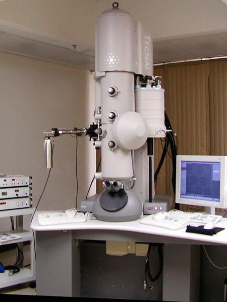
Electron microscope
The history of the development of the electron microscope began in 1931, when a certain R. Rudenberg received a patent for the first transmission electron microscope. Then, in the 40s of the last century, scanning electron microscopes appeared, which reached their technical perfection already in the 60s of the last century. They formed an image of the object due to the sequential movement of a small-section electronic probe over the object.
How does an electron microscope work? At the heart of its work is a directed beam of electrons accelerated in an electric field and displays the image on special magnetic lenses, this electron beam is much smaller than the wavelength of visible light. All this makes it possible to increase the power of an electron microscope and its resolution by 1000-10,000 times in comparison with a traditional light microscope. This is the main advantage of the electron microscope.

This is what a modern electron microscope looks like.
Laser microscope
A laser microscope is an improved version of an electron microscope, its operation is based on a laser beam that allows the scientist's gaze to observe living tissues at an even greater depth.
X-ray microscope
X-ray microscopes are used to examine very small objects with dimensions comparable to the dimensions of an X-ray wave. Their work is based on electromagnetic radiation with a wavelength of 0.01 to 1 nanometer.
Microscope device
The design of a microscope depends on its type, of course, an electron microscope will differ in its structure from a light optical microscope or from an X-ray microscope. In this article, we will consider the structure of a conventional modern optical microscope, which is the most popular among both amateurs and professionals, since they can be used to solve many simple research problems.
So, first of all, in a microscope, you can distinguish the optical and mechanical parts. The optical part includes:
- The eyepiece is the part of the microscope that is directly connected to the eyes of the observer. In the very first microscopes, it consisted of a single lens; the design of the eyepiece in modern microscopes, of course, is somewhat more complicated.
- The objective is practically the most important part of the microscope, as it is the objective that provides the main magnification.
- Illuminator - responsible for the flow of light onto the object under study.
- Aperture - adjusts the strength of the luminous flux entering the object under study.
The mechanical part of the microscope consists of such important parts as:
- A tube, it is a tube that contains an eyepiece. The tube must be strong and not deformed, otherwise the optical properties of the microscope will suffer.
- The base, it provides stability of the microscope during operation. It is on it that the tube, capacitor holder, focusing knobs and other parts of the microscope are attached.
- Revolving head - used for quick change of objectives, it is absent in cheap models of microscopes.
- The subject table is the place on which the investigated object or objects are placed.

And here the picture shows a more detailed structure of the microscope.
Rules for working with a microscope
- It is necessary to work with the microscope while sitting;
- Before work, the microscope must be checked and wiped off dust with a soft cloth;
- Place the microscope in front of you a little to the left;
- It is worth starting work with a small increase;
- Set the lighting in the field of view of the microscope using an electric light or mirror. Looking through the eyepiece with one eye and using a mirror with a concave side, direct the light from the window into the lens, and then illuminate the field of view as much and evenly as possible. If the microscope is equipped with an illuminator, then connect the microscope to a power source, turn on the lamp and set the required brightness of combustion;
- Place the micropreparation on the stage so that the object under study is under the objective. Looking from the side, lower the lens using the macroscrew until the distance between the lower lens of the objective and the micropreparation becomes 4-5 mm;
- Moving the specimen with your hand, find the right place, place it in the center of the microscope field of view;
- To study an object at high magnification, you first need to place the selected area in the center of the microscope's field of view at low magnification. Then change the objective to 40x by turning the revolver so that it is in the working position. Achieve a good image of the object using the micrometer screw. There are two dashes on the box of the micrometer mechanism, and on the micrometer screw there is a point that should always be between the lines. If it goes beyond their limits, it must be returned to its normal position. If this rule is not followed, the micrometer screw may stop working;
- Upon completion of work with a high magnification, set a low magnification, raise the lens, remove the specimen from the work table, wipe all parts of the microscope with a clean napkin, cover it with a plastic bag and put it in a cabinet.

When writing the article, I tried to make it as interesting, useful and high-quality as possible. I would be grateful for any feedback and constructive criticism in the form of comments to the article. Also, you can write your wish / question / suggestion to my mail [email protected] or Facebook, sincerely the author.


 The wealth of a language is determined not only by wealth")
